In the first step to access the zinc complex, ring closure occurred in the reaction of phenylhydrazine and ethyl-2-(ethoxymethylene)-4,4,4-trifluoro-3-oxobutanoate in the presence of triethylamine, leading to intermediate 128. Hydrazide compound (2) was obtained by the reaction of hydrazine hydrate with the ester compound. The synthesis of the ligand (IMP) was carried out via imine bond formation of the hydrazide compound with 2-Pyridinecarboxaldehyde in the presence of a catalyst (AcOH). The zinc complex (IMP-Zn) was prepared with the addition of ZnCl2 to the methanolic ligand solution (Scheme 1). Attempts were made to grow single crystals of the Zn(II) complex suitable for X-ray diffraction using slow evaporation and diffusion methods in various solvent systems (methanol, ethanol, DMF, and mixed solvents). However, these trials yielded either microcrystalline solids or crystals of insufficient quality for SC-XRD analysis. This is attributed to the flexible hydrazone framework and the labile coordination nature of Zn(II), which often limits the formation of well-ordered single crystals. Therefore, the structural assignment of the complex was established based on complementary spectroscopic, analytical, and computational techniques.

Synthetic pathway for the target complex.
FTIR analysis
Many reports with interesting biological applications have adopted the analysis of vibrational spectra of hydrazone derivatives as well as their metal complexes had recently appeared29,30,31,32,33. Here, we have carried out DFT computational comparative vibrational studies of the current IMP ligand and its IMP-Zn complex in order to precisely assign the positions of the important functional groups that usually serve donor groups to the metal species. The investigated vibration wavenumbers were computed in the frequency range 4000–400 cm-1. Calculation of the wavenumbers was based on the symmetry of a C1 point group. The experimental and calculated IR spectra of IMP ligand and its zinc complex are given in the supplementary file (Fig. S1). Table 1 gives a summary of the most characteristic experimental wavenumbers versus the calculated values. The objective of the assignment was to assess the activity in IR and to compare the experimental and calculated intensities. The experimental and calculated values of νNH of IMP ligand were detected at comparable ranges (Table 1). The corresponding stretching values of NH group in the experimental and calculated IR spectra of IMP-Zn complex were shifted to lower frequencies due to complex formation. In addition, the experimental νC = O (C = O stretching coupled with the NH group, amide I) and νC = N stretching frequencies of the ligand were observed at 1663 and 1652 cm-1 (calculated values were 1706 and 1674 cm-1). These stretches also displayed shifts to lower frequencies in the spectra of the IMP-Zn complex. The observed small value of the νC = O could be due to the presence of the trifluoromethyl-pyrazole moiety, which might alter the electronic structure of the whole molecule. The bands in the range of 1500–1110 cm-1 were attributed to the CH and NH bending. It is worth mentioning that some vibrations were coupled with other coordinates. Furthermore, two experimental N–N stretching vibrations for the IMP hydrazone and pyrazole parts were detected at 1167 and 1179 cm-1, while the calculated value was 1158 and 1180 cm-1 34,35,36. Again, these bands were also shifted to lower values on complexation with the zinc ion. Previous investigations of some hydrazone ligands and their metal complexes revealed comparable values for these stretches29. For the assignment of other modes, the in-plane and out-of-plane ring bending were found at lower frequencies. The most characteristic ones were the in phase γCH and γC = O out-of-plane modes, which were observed with strong comparable intensities at around 740 cm-1. Furthermore, asymmetric and symmetric stretching of the Cl-Zn-Cl bonds were detected in the calculated IR spectrum at 340 and 274 cm-1, respectively. Previous investigation of the calculated IR spectrum of the [Au(pyrazinecarboxamide)(Cl)2] complex showed that the νasCl-Au-Cl and νsCl-Au-Cl occurred at 373 and 353 cm-1, respectively. These values were confirmed by Raman analysis37,38.
UV–Vis analysis
The UV–Vis spectrum of the IMP-Zn complex was measured in DMF solution at a concentration of 10 µM. The free ligand IMP spectra demonstrated a band at 296 nm, which corresponds to the intraligand π → π* transition according to the log ε of 4.65. The π → π* transition absorption band was displaced to 292 nm in the IMP-Zn spectra, indicating the coordination of the zinc(II) ions to the hydrazone ligand. The formation of the complex is verified too by the presence of a band in 372 nm, characteristic of ligand–metal charge transfer (LMCT) transition (Table 2). Additionally, conductivity study was performed to determine the nature of the complex in solution. The measurement was carried out using a digital conductivity meter equipped with a cell constant appropriate for low-concentration solutions was prepared in DMF and the measurement was taken at 25 °C to ensure accurate results. The molar conductivity of IMP-Zn was found to be 9.15 Ω−1.cm2.mol−1. Such a low molar conductance indicates that the complex behaves as a non-electrolyte under these conditions, consistent with chloride ligands being coordinated to the zinc center rather than existing as counter-ions in solution36. The solution stability of IMP-Zn in DMF was evaluated by time-dependent UV–Vis spectroscopy. A 10 µM solution of IMP-Zn in DMF was stored at 25 °C in the dark and spectra were recorded at regular time intervals over 48 h (0–48 h). The characteristic absorption bands assigned to the intraligand transition and LMCT band remained unchanged in position and intensity, indicating that the complex is stable in DMF over the examined period (Fig. S7). This conclusion is supported by FT-IR, NMR, elemental analysis and mass spectrometry data as well as DFT results, which together indicate a neutral/coordinated complex formulation. The bathochromic shift observed in the IMP-Zn complex indicates reduced HOMO–LUMO energy gap and enhanced charge transfer ability due to coordination, which is in agreement with the DFT results and consistent with the higher biological reactivity of the complex compared to the free ligand39.
NMR analysis
To understand the solution-phase behavior of both ligand and complex and to verify their consistency with the solid-state structure, 1H NMR analyses of IMP and IMP-Zn were conducted in DMSO-d6 medium. Considering 1H NMR spectra of the ligand, the pyrazole C-3 proton resonated at 7.42 ppm. The aromatic protons in the phenyl ring attached to pyrazole ring were found at 7.59 ppm as a singlet. The pyridine ring protons resonated in the range of 7.74 and 8.36 ppm, and imine proton (N = CH) was 8.60 ppm as a doublet. The hydrazone NH proton in the ligand resonated at 12.20 ppm. In the 13C NMR spectra of the ligand, the pyrazole C-3 carbon was found at 140.28 ppm. It was also seen that the C = O carbon peak was found at 163.17 ppm in 13C NMR (APT). The pyrazole C-5 carbon resonated at 139.12 ppm, while the imine carbon was observed at 145.54 ppm. The observations supported that the ligand is in keto form in solution-phase. When examining the NMR spectra of the synthesized zinc complex, it was observed that the most significant changes are the disappearance of the NH proton in 1H NMR and the observation of the C = O peak at 171.01 ppm in 13C NMR. It was observed that, upon examination of the peaks, the imine proton was found at 8.71 ppm, while the pyrazole C-3 proton was found at 7.71 ppm. Significant shifts have been observed in the aromatic ring protons, with the most pronounced shift occurring in the CH proton adjacent to nitrogen in the pyridine ring40. The proton is observed at 8.34 ppm (doublet) in the ligand, while it is observed at 8.15 ppm (singlet) in the complex. Similarly, within the 13C NMR spectrum of the IMP-Zn, the pyrazole C-5 carbon peak resonated at 139.85 ppm and the imine carbon at 147.65 ppm. The pyrazole C-3 carbon was detected at 140.51 ppm. These shifts are attributed to changes in the electronic density of the hydrazone ligand upon coordination with the zinc(II) atom. As a result, the NMR data suggests that IMP-Zn exhibits dynamic solution behavior in DMSO, which may involve partial dissociation or ligand exchange, a common feature of Zn(II) Schiff-base complexes in polar media (Fig. S2). Overall, based on spectroscopic analysis, conductivity measurements, and DFT optimization, IMP-Zn can be best described as a penta-coordinated Zn(II) complex; formation of a bis-ligated octahedral species was not supported under the present synthetic conditions.
Thermal properties
The thermal stability and decomposition pathways of IMP and its zinc(II) complex (IMP-Zn) were investigated by thermogravimetric analysis (TGA) (Fig. 1). The free ligand displayed a single major weight-loss event between 165 and 380 °C, corresponding to the decomposition of the hydrazone framework, with an overall mass loss of 84.48%. In contrast, the thermal decomposition profile of IMP-Zn exhibited two distinct steps, indicating higher stability due to metal coordination. The first stage (280–525 °C) involved a 59.96% mass loss, assignable to the removal of coordinated organic moieties bound to the Zn(II) center. The second degradation step (610–890 °C) accounted for an additional 22.35% loss and was attributed to the decomposition of the more stable aromatic backbone. The final residue (17.68%) matched well with the theoretical value of ZnO, confirming the identity of the metal content37,38. The data confirmed that coordination with Zn(II) enhances the thermal robustness of the hydrazone ligand, shifting decomposition to higher temperatures and leading to a more gradual degradation profile. This enhanced stability is consistent with the chelation effect and supports the structural assignment of IMP-Zn.

TGA analysis of ligand and zinc complex.
SEM analysis
A surface morphology examination of the synthesized ligand and complex was conducted via SEM analysis. The resulting micrographs are presented in Fig. 2. The micrograph of IMP is displayed in Fig. 2A; the image demonstrates an irregular surface morphology with aggregates of varying sizes and shapes. The rough and fragmented appearance of the surface suggests the presence of porosity, which is indicative of a non-uniform texture. Conversely, the micrographs of zinc complex (Fig. 2B) exhibited irregular broken rock-type morphology.
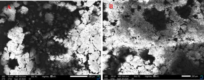
Scanning electron micrographs of (A) IMP and (B) IMP-Zn.
In silico calculationsStereochemistry, natural bond orbital and global reactivity descriptors calculations
The stereochemistry of the energy-minimized geometries, energies and global reactivity descriptors for the pyrazole-carbohydrazide derivative and its zinc complex were computed by using the DFT method implemented in Gaussian 09w software. The global reactivity descriptors were calculated using the following equations:
$$\Delta E \, = \, – \, (E_{HOMO} ) \, + \left( {E_{LUMO} } \right)$$
$$IP = – E_{HOMO} ,EA = – E_{LUMO}$$
$$V = – \chi = – {1}/{2}\left( {IP + EA} \right)$$
$$\eta = {\raise0.5ex\hbox{$\scriptstyle 1$} \kern-0.1em/\kern-0.15em \lower0.25ex\hbox{$\scriptstyle 2$}}\left( {IP – EA} \right)$$
$$\omega = (V^{{2}} /{2}\eta )$$
Frontier molecular orbitals—specifically the highest occupied molecular orbital (HOMO) and the lowest unoccupied molecular orbital (LUMO)—are frequently analyzed to gain insight into the optical and electronic properties of compounds. These orbitals also provide valuable information for assessing various intramolecular charge transfer (CT) mechanisms, as well as the chemical reactivity and stability of the molecules under investigation32,41,42. The energy level of the HOMO is associated with the molecule’s ability to donate electrons and is typically described by the ionization potential. Conversely, the LUMO energy level reflects the molecule’s potential to accept electrons and corresponds to the electron affinity. The energy difference between HOMO and LUMO, referred to as the energy gap (ΔE), plays a critical role in determining molecular properties. A smaller ΔE suggests efficient intramolecular charge transfer from donor to acceptor sites, indicating higher reactivity and lower stability. In contrast, a larger ΔE is indicative of reduced reactivity and greater molecular stability. IR and 1^11H-NMR analyses of the synthesized ligand confirmed the presence of keto-enol tautomerism. Consequently, the lowest-energy optimized geometries for both the keto and enol tautomers were calculated, focusing on the spatial orientation of key functional groups, including C=O, N–NH, C=N–N=C–OH, as well as pyridyl and pyrazole units. Fig. S4 presents the optimized molecular structures alongside the Mulliken charge distribution maps for both tautomeric forms. The energy-minimized keto and enol forms in order to compare the energy and the different structural arrangements, which can help in deducing the best coordination site of the ligand as well as to correlate with the spectroscopic data such as IR spectra. The keto and enol forms exhibited energies of 32.93 and 37.59 kcal/mol, respectively, and displayed distinct structural features. In both forms, the molecule adopted a non-planar, asymmetrical geometry classified under the C1 point group. The pyridine ring, C=N–NH–C=O (or C=N–N=C–OH), and the pyrazole ring were coplanar, with a dihedral angle—such as N14–N15–C16–C17—measured at approximately 179.9° in both tautomers. However, the phenyl ring attached to the N7 atom of the pyrazole was tilted, showing dihedral angles of 38.6° and 40.9° for the keto and enol forms, respectively. Notably, in the optimized enol structure, the hydroxyl hydrogen was oriented toward the N15 atom of the N–N group, forming an intramolecular hydrogen bond with a distance of 1.97 Å (Fig. 3). The bond lengths and bond angles of the rest of the molecule were in normal values observed before29,43,44. The charge allocations on the different atoms of the two isomers were calculated using the Mulliken method, Fig. S4. From the charge distribution findings, for both isomers, the pyrazole nitrogen attached to the phenyl group (N7) had the highest negative charge (− 0.653 and − 0.659, for the keto- and enol-forms, respectively). However, according to its crowding site, this donor atom was not available for coordination. On the other hand, the carbonyl oxygen (O13), the nitrogen atoms of N–H (N14) and pyridyl (N22) groups of the keto-form had the highest negative charges (− 0.420, − 0.545 and − 0.389, respectively). In the case of the enol-form, the calculated charge on the hydroxyl oxygen (O13), C=N (N14) and pyridyl (N22) groups had the highest negative values (-0.563, -0.316 and -0.382, respectively), Fig. 3.

The geometrically optimized structures of the keto- and enol-forms of IMP ligand.
The lowest energy-minimized orientation of the IMP-Zn was computed by focusing on the arrangement of the important functional groups of the coordinated ligand as well as the zinc coordination core. Figure 4 displays the geometrical optimization structure along with some bond lengths and bond angles around the zinc species. The Mulliken charge distribution map of the complex is given in Fig. S5. All the other bond lengths and bond angles of the investigated complexes were in normal values as those observed before29,43,44. The ligand acted as a tridentate and coordinated with the metal ion from the carbonyl oxygen and the C=N and pyridyl nitrogen atoms. The coordination sphere of the zinc ion was completed by two coordinated chloride species to give a distorted trigonal bipyramidal structure36. The distorted structure stemmed from the deviated bond length and bond angle values of a regular trigonal bipyramidal arrangement. According to the obtained angles, the equatorial base of the trigonal bipyramid consisted of N15 and the two chloride species, while the axial position was occupied by the pyridyl nitrogen (N23) and carbonyl oxygen (O13) (Fig. 4). The Mulliken charge distribution on the atoms of the complex displayed dramatic changes relative to those of the free ligand, Fig. S5. Obviously, the charge on the donor atoms was reduced due to the complex formation. Moreover, the electrostatic potential (ESP) maps of the two isomers of the ligand and the Zn complex (Fig. 5) provided important information about the charge distributions within the compounds. These plots distinguished the reactive zones of the investigated compounds by displaying both the electrophilic and the nucleophilic attack regions. The electrostatic potential maps are also effective representations that give useful information in the biological fields45. The ESP map determines the positive (blue color) and negative (red color) charged electrostatic potential. The color scale of the two isomers of the IMP ranged from -0.07951 Hartree (− 208.75 kJ/mol) to + 0.07951 Hartree (+ 208.75 kJ/mol) for the keto-form, while it ranged from − 0.06918 Hartree (− 181.63 kJ/mol) to + 0.06918 Hartree (+ 181.63 kJ/mol) for the enol-form. In the case of IMP-Zn, the color scale ranged from − 0.09695 Hartree (− 254.54 kJ/mol) to + 0.09695 Hartree (+ 254.54 kJ/mol). High negative potential red zones indicated the electrophilic attack sites, while high positive potential blue sections designated appropriate centers for nucleophilic attack. It is clear from the ligand map that the oxygen and nitrogen donor atoms were the most susceptible zones for the electrophilic attack of the zinc ion. For the IMP-Zn the two coordinated Cl groups showed the high negative electrostatic potential.

The geometrically optimized structure of the IMP-Zn complex.

The electrostatic potential (ESP) maps for the ligand isomers and the IMP-Zn.
Natural Bond Orbital (NBO) is a useful approach to determine the nature of electrophilic and nucleophilic hyperconjugative interactions, intramolecular charge delocalization and other bonding interactions that involves in the electronic transitions46. This includes the transfer of charge from occupied orbitals, defined as Lewis-type NBOs, to unoccupied orbitals, as non-Lewis-type NBOs). Tables S1 and S2 give complete data sets for both ligand and its zinc complex. The second-order perturbation theory was applied to determine the stabilization energy E(2), which quantifies the donor(i)-acceptor(j) interactions, i.e., the delocalization of electron density from a filled donor orbital to an empty acceptor orbital (i → j)47. The E(2) was calculated using the formula:
$$E\left( 2 \right) = – {\text{q}}_{{\text{i}}} \left| {{\text{F}}_{{{\text{ij}}}} } \right|^{{2}} /\left( {\varepsilon_{{\text{j}}} – \varepsilon_{{\text{i}}} } \right)$$
qi= the occupancy of the donor orbital,
Fii= the off-diagonal Fock matrix element between the donor and acceptor orbitals,
εj= the energy of the non-Lewis NBO,
εi;= the energy of the occupied Lewis NBO.
Overlapping of the orbitals σ → σ*, π → π*, LP → σ*, and LP → π* caused hyper-conjugation phenomenon. The σ → σ* transitions were more detected among these transitions, and they were possible by the interaction between the electron-rich and electron-poor components. The π → π* transitions provided support to the π-conjugated structures48,49. On the other hand, the accretion of natural charges on the individual atoms coordinated with the zinc ion before and after complexation, the natural population of the electrons of the metal ion in the core, valence and Rydberg sub‐shells and natural electronic configuration of Zn2+ in the coordination globe of the complex are given in Tables 3 and 4. The most electronegative charges are accumulated on the nitrogen and oxygen atoms of the ligand and the complex. The charge allocations on the different atoms of the ligand and its zinc complex were calculated using both Mulliken and natural bond orbital methods, (Tables S1 and S2, Figs. S4 and S5). The charge distribution findings of both two methods showed some discrepancies. The NBO method showed higher negative values of the coordinated donor atoms relative to the results of Mulliken method. The pyrazole nitrogen attached to the phenyl group (N7) and its adjacent nitrogen (N8) as well as the NH atom (N14) are not available for coordination according to their crowding site. The electronegative atoms in the coordination sphere tend to provide electrons to the central metal ions. The most electropositive charges accrued on the Zn and the carbon atom of CF3 (C26) species (1.0744 and 1.1105). Obviously, C26 has the highest positive charge due to the attached three fluoride atoms. In case of Zn, it is more likely to receive electrons from the ligand. This is clearly indicated from the natural electronic configuration (Table 3). Back donation from the zinc orbitals to the coordinated chloro ligands is also confirmed from such electronic configuration ([core] 4S0.40 3d9.99 4p0.52 5p0.01).
The global chemical reactivities of the two reported compounds, such as the energy gap (ΔE), chemical potential (V), ionization potential (I), electron affinity (A), electronegativity (Χ), chemical softness (S), chemical hardness (η) and electrophilicity index (ω) as well as the total energy and dipole moment (DM) values were estimated from the energy values of the HOMO and LUMO orbitals (Table 5)37,50. The HOMO and LUMO orbitals of the keto-form of the ligand and its Zn derivative are illustrated in Fig. 6. The electron cloud of the frontier orbitals of the IMP was mostly localized over the pyridine ring and C = N–NH-C = O parts. On the other hand, the distribution of the HOMO orbital of the IMP-Zn was localized on the zinc and the two chloride ions, while the LUMO orbital was located on the pyridine ring and C = N–NH-C = O moieties as well as the two nitrogen atoms of the pyrazole ring. The energy gap (ΔE) of the two compounds was 4.45 and 2.38 eV for the ligand and the complex, respectively. This means that the charge transfer within the molecule (from HOMO to LUMO orbital) is more responsive in the complex than in the ligand. This in turn reflects that the IMP-Zn had higher chemical reactivity and lower stability relative to the free ligand. In addition, the IMP-Zn was characterized by lower IP and higher EA relative to those of ligands.

The HOMO and LUMO orbitals and the energy gap (ΔE) values of the keto-form of the ligand isomers and the IMP-Zn.
Furthermore, the observed small values of the chemical hardness and chemical softness for the zinc complex versus those of the ligand indicated the ability of CT within the molecule and confirmed the expected higher chemical reactivity and lower stability of the complex. Furthermore, the smaller value of ΔE for the zinc complex and its linear relationship with both chemical hardness and chemical softness values confirmed the preference for better biological activity of the complex over the IMP ligand. This conclusion can also be stemmed from the higher value of the electrophilicity index of the IMP-Zn complex (Table 5). The electrophilicity index covers the electronegativity of the electron acceptor and its chemical hardness and acts as a resistance to the electronic charge exchange32,41.
Molecular docking investigations
The molecular docking study is a useful method for predicting the different interactions between a synthesized guest molecule and a selected host macromolecular biological target51. The molecular docking process typically involves the evaluation of the conformation poses that are generated from the binding zones of the biological targets to accommodate a hydrophobic inhibitor. Furthermore, it has been demonstrated to deduce the manner in which a docked compound is accommodated within the minor grooves of a macromolecular receptor. Conversely, bioactive nitrogen heterocyclic derivatives, including pyrazoles, pyridines, and pyrazines, represent significant components in drug design, exhibiting a diverse array of biological activities and being extensively utilized in a variety of clinically tested drugs52,53. So, the study of molecular docking of these derivatives surely helps in assorting and exploring new drugs containing such important derivatives. Therefore, the molecular docking studies of the reported IMP and IMP-Zn were evaluated using three macromolecular receptors, namely, a kinase extract from the epidermal growth factor receptor (PDB: 1M17), a protein extract from cyclin-dependent kinase inhibitors (PDB ID: 6GUE) and a protein extract from cytochrome P450 (PDB: 3RUK). These protein receptors were chosen to correlate the docking findings with experimental cytotoxicity data. Furthermore, the study was executed to declare the different interaction styles and to identify the various plausible binding poses and energies. The binding score values, hydrophobic interactions and the different types of hydrogen bonding were the main factors that were used to select the best molecular conformation pose between target and inhibitor. The docking methodology was validated to ensure that the used procedures and input parameters were reliable and gave accurate predictions for the ligand-receptor interactions. Thus, the validation process was built on many factors such as RMSD (root mean square deviation), scoring function values and the numbers and types of bonding interactions. The RMSD value is the important key parameter for deducing the accuracy of the docking procedure. It determines the average displacement between the atomic positions of the docked ligand and its original conformation. The method is considered optimal with high precision when the RMSD value is less than 2 Å. However, the method is considered acceptable and reasonably reliable if the RMSD value is between 2 and 3 Å54. Furthermore, the reasonably high negative score function values for the current ligand and the complex towards the selected receptors concluded their high binding affinity (Table 6). Figures 7, 8 and 9 display the two- and three-dimensional plots of the interacting ligand and its corresponding Zn complex with the docked protein receptors. The molecular docking data like score functions, RMSD values, types and energies of binding interactions are given in Table 6. The RMSD values obtained from the molecular docking operations with the selected receptors ranged from 0.82 and 2.09 Å, concluding that the docking processes were adequately acceptable and reliable. Moreover, the docking findings revealed that the hydrophobic ligand (IMP and IMP-Zn) exposure was the major interaction type. It is worth mentioning that the polar parts of the compounds, such as F, Cl, N and O atoms, along with the aromatic moieties had the highest hydrophobic retardation55,56.

The two- and three-dimension representations of the interactions of the IMP and IMP-Zn with 1M17 target.

The two- and three-dimensional representations of the interactions of the IMP and IMP-Zn with 6GUE target.

The two- and three-dimension representations of the interactions of the IMP and IMP-Zn with 3RUK target.
The molecular docking of the free ligand and its zinc derivative versus 1M17 receptor showed binding interactions with specific residues of the following amino acids: glutamic acid (Glu), arginine (Arg), aspartic acid (Asp), asparagine (Asn), tyrosine (Tyr), leucine (Leu), glutamine (Gln), lysine (Lys), histidine (His), and alanine (Ala) (Fig. 7). For the free ligand, the amino acid residues Lys 733 and Lys 836 exhibited π-Hbinding interaction with the phenyl ring of the ligand. In the case of the zinc complex, the amino acid residue Arg 817 moiety displayed a basic-backbone hydrogen donor. On the other hand, the Asp 831 exhibited ionic interaction with one of the coordinated chloride of the complex (Table 6).
Furthermore, the molecular docking of the two reported compounds with the 6GUE target displayed binding interactions to similar residues of the amino acids as in the case of 1M17 receptor (Fig. 8). Two different π-interactions were noted between the IMP and the Gln 131 and Lys 129 residues. The former interaction was of the π-H type from the π-electrons of the phenyl group while the latter was π-cation interaction from the π-electrons of the pyridyl group. In addition, the docking data exhibited polar hydrogen bonding interaction, with receptor exposure, between the carbonyl oxygen of the ligand and the tyrosine residue Tyr 15. The IMP-Zn showed π-H interaction from the phenyl ring to the His 84A residue. It also demonstrated a basic sidechain H-donor between one of the coordinated Cl and the amino acid part Lys 202B. Further, the complex underwent ionic interaction between the two nitrogen atoms of the N–NH group and the Glu 138A residue, Table 2. On the other side, the 3RUK receptor interacted with the ligand from residues of the amino acids: arginine (Arg), serine (Ser), valine (Val), glycine (Gly) and methionine (Met), while it interacted with the zinc complex from the amino acid parts: proline (Pro), aspartic acid (Asp), glutamic acid (Glu) and valine (Val). The ligand displayed two different π-H interactions (pyridyl ring-Ser 65 residue and pyrazole ring-Arg 67 residue). The zinc complex also showed two π-H interactions between the phenyl ring with Val 197 residue, and the pyrazole ring with Pro 193 residue. In addition, the zinc complex exerted ionic interactions between the pyridyl nitrogen, carbonyl oxygen, and the two nitrogen atoms of the N–NH group versus the amino acid residue Asp 192. The experimental cytotoxic effects of the IMP ligand and its IMP-Zn complex on the tested HCT116 cell line are consistent with the molecular docking of the two receptors 1M17 and 3RUK, where the complex displayed higher binding affinity (higher score values) relative to the free ligand. Following the findings of this study, Ghasemi et al. have showed that mixed-ligand Schiff base complexes exhibit cytotoxic effects on HCT116 cells and displayed a strong binding affinity for 3RUK protein, as determined by molecular docking studies57. In a separate study conducted on the HCT116 cell line, molecular docking studies demonstrated a strong correlation with in vitro findings, thereby suggesting the potential of organoselenium complexes as colorectal cancer inhibitors via interaction with cytochrome P45058. In conclusion, the order of the binding affinity towards the two receptors, 1M17 and 3RUK, was as follows: IMP-Zn > IMP. However, the interaction of the two compounds with the 6GUE receptor was the reverse, i.e., IMP > IMP-Zn. This phenomenon can be attributed to the distinct arrangement of amino acid residues, in conjunction with the availability of minor grooves and their respective dimensions.
As can be seen from Table 6 for all the tested three receptors, the score function (S) values for the zinc complex are higher than those of the IMP ligand that means higher binding affinity of the complex towards the receptors. This is consistent with and correlated to the experimental cytotoxic finding where the IMP-Zn complex displays lower IC50 relative to the IMP ligand itself (Table 7). Furthermore, the docking results are harmonious with the reactivity descriptor data (Table 5). It was found that the IMP-Zn complex has small values for the ΔE, chemical hardness and chemical softness along with higher value of the electrophilicity index, which confirmed the expected higher chemical reactivity and lower stability.
Anticancer activityCytotoxic effect of synthesized IMP and IMP-Zn
The effect of IMP and IMP-Zn on cell viability of HCT116 and HEK293 cell lines was analyzed using the resazurin viability assay. HCT116 and HEK293 cells were treated with IMP-Zn and IMP at concentrations ranging from 10 to 50 μM, and cell viability was assessed at 24-h (h) intervals. Untreated cells were used as a negative control. The complexes were dissolved in DMF to prepare the main stock solution and diluted with DMEM to the desired concentration. The final concentration of DMF did not exceed 0.1%. As was stated in the preceding report59, the DMF has been demonstrated to exhibit no cytotoxic effects on cell growth (Figure S6).
IMP-Zn and IMP showed a decrease in cell viability of HCT116 cells in a time and dose-dependent manner (Fig. 10). A significant decrease in viable cells was observed at all three-time points with increasing concentration in both complexes compared to untreated controls. This finding indicated that the cytotoxic effect of IMP-Zn is significantly higher compared to IMP at these dosages.

Resazurin cell viability analysis of colon cancer cell line HCT116 treated with IMP-Zn and IMP. The cytotoxic effect of (A) IMP-Zn and (B) IMP on HCT116 cell line was investigated at concentrations ranging from 0 to 50 μM for 24, 48, and 72 h compared to untreated cells. Data represent mean (n = 3) ± SD (nsp ≥ 0.05, *P < 0.05, **P < 0.01, ***P < 0.001, ****P < 0.0001).
The cytotoxic efficacy of the IMP-Zn, and IMP complexes in the HCT116 cell line was determined by the median growth inhibitory concentration (IC50), which requires a 50% cytotoxic effect against cancer cells. The IC50 values for IMP-Zn were 28.62 μM, 24.56 μM, and 24.28 μM and the IC50 values for IMP were 54.61 μM, 37.58 μM, and 31.90 μM at 24, 48, and 72 h, respectively. Thus, the IC50 value of IMP-Zn was found to be lower than that of IMP at all three time points (Table 7). Our results were consistent with studies that reported zinc Schiff base complexes showed higher cytotoxic effects on cancer cells compared to their free ligands60. Therefore, the higher cytotoxic effect of the Zn (II) complex on human colon cancer cell viability at lower doses was suggested to be related to its stronger DNA binding ability60,61,62.
Comparing the previous studies, recent reports have highlighted the anticancer potential of Zn(II) Schiff base and hydrazone complexes, but most studies have focused on either antimicrobial activity or enzyme inhibition rather than direct cytotoxic evaluation in colorectal carcinoma models. For instance, Santiago et al. reported hydrazone-derived Zn(II) complexes with moderate antibacterial effects and limited cytotoxic potential (IC50 > 60 μM in most tested cancer cell lines)35. Similarly, Dasgupta et al. described acyl-hydrazone Zn(II) complexes with phosphatase inhibition and modest anticancer activity, but with higher IC50 values compared to our complex36. In contrast, our IMP-Zn complex exhibited significantly enhanced cytotoxicity toward HCT-116 cells with an IC50 of 24.56 μM at 48 h, underscoring its superior potency relative to structurally related Zn(II) derivatives.
Furthermore, HEK293 cell line is used as normal cell line to compare the cytotoxic effect of IMP-Zn and its free ligand. The exposure of HEK293 cells to IMP-Zn and IMP for 48 h had no significant effect on cell viability up to a concentration of 50 μM (Figure S4). Cytotoxicity studies on colon cancer show that HEK293 cell line could be selected as normal cell line63. Our cell viability results showed that HCT116 cancer cells were significantly more sensitive to IMP-Zn and its free ligand compared to HEK293 cells.
Notably, pyrazole-based Zn(II) complexes have been reported to show interesting photophysical or antioxidant properties22,23,24, yet their anticancer potential remains largely unexplored. Our findings expand this scope by demonstrating that the incorporation of a pyrazole-hydrazone scaffold into a Zn(II) coordination sphere not only stabilizes the ligand but also enhances biological reactivity against colon carcinoma cells. Taking together, these comparisons emphasize that our work does not simply reiterate previous studies but introduces a novel pyrazole-hydrazone Zn(II) scaffold with demonstrably higher anticancer activity, thereby contributing to the growing field of zinc-based metallodrugs.
IMP-Zn decreases colony formation of HCT116 cells
To investigate the effect of IMP-Zn on colony formation in HCT116 cells, cells were incubated with 10 μM, 20 μM, and 24.56 μM IMP-Zn for 48 h, fresh medium was added at the end of 48 h, and cells were allowed to colony formation for 14 days. Colony diameters and numbers were calculated compared to the control cells. As shown in Fig. 11, colony numbers and diameters were significantly decreased in cells treated with increasing doses of IMP-Zn (10, 20, 24.56 μM) compared to the control group, which is parallel in our cell viability results (Fig. 10). Images of the colonies show less in number for the IMP-Zn treated groups compared to the control group (Fig. 11A). Colony diameters were calculated as 65.55 μm, 41.16.98 μm, 27.06 μm, and 21,93 μm for control, and IMP-Zn-treated groups (10, 20, and 24.56 μM), respectively (Fig. 11B). Colony numbers were 87, 70, 39, and 9 for the control, and IMP-Zn-treated groups (10, 20, and 24.56 μM), respectively (Fig. 11C).

Colonization of HCT116 cells for 14 days after 48 h treatment with IMP-Zn (10, 20 and 24.56 μM). (A) Representative well images of colonies (left panel) and colonies of HCT-116 cells taken with a 10 × objective (right panel). (B) Graphical representation of colony numbers and (C) diameters (μm) for control and IMP-Zn-treated HCT116 cells. Data represent mean (n = 3) ± SD (nsp ≥ 0.05, *P < 0.05, and **P < 0.01). Scale bar: 20 μm.
Effects of IMP-Zn and IMP on Cell Cycle
The effects of IMP-Zn and IMP on cell cycle progression in HCT116 cells were investigated using flow cytometry with PI staining. Cell populations in SubG0, G0/G1, S, and G2/M phases were analyzed based on their DNA content in control cells and in cells treated with 24.56 μM IMP-Zn and IMP for 48 h. As shown in Fig. 12, the percentage of HCT116 cells at Sub G0 phase were 5.86%, 20.51%, and 10.82% for the control, IMP-Zn, and IMP treatment groups, respectively. Thus, treatment of HCT116 cells with IMP-Zn resulted in a significant increase in the SubG0 phase. SubG0 represents cell population with reduced DNA stainability that is most likely due to DNA fragmentation64. An anticancer study on zinc Schiff base complex pointed out that increased SubG0 population represents DNA fragmentation in cells undergoing apoptosis65. Thus, the increased Sub-G0 phase in IMP-Zn treated HCT-116 (Fig. 12) might be an indication of apoptosis. The percentage of S phase cell population were 12.04%, 14.28%, and 13.95% for the control, IMP-Zn and IMP, respectively, and 34,50%, 32,88% and 34,61% in G2/M phase, respectively (Table S3).

The cell cycle progression of HCT116 cells was analyzed following treatment with 24.56 μM IMP-Zn and IMP for 48 h compared to untreated control cells. (A) Representative histograms and (B) graphical representation of cell cycle distribution of SubG0, G0/G1, S, and G2/M phases.
Taking together, these comparisons emphasize that our work does not simply reiterate previous studies but introduces a novel pyrazole-hydrazone Zn(II) scaffold with demonstrably higher anticancer activity, thereby contributing to the growing field of zinc-based metallodrugs.