
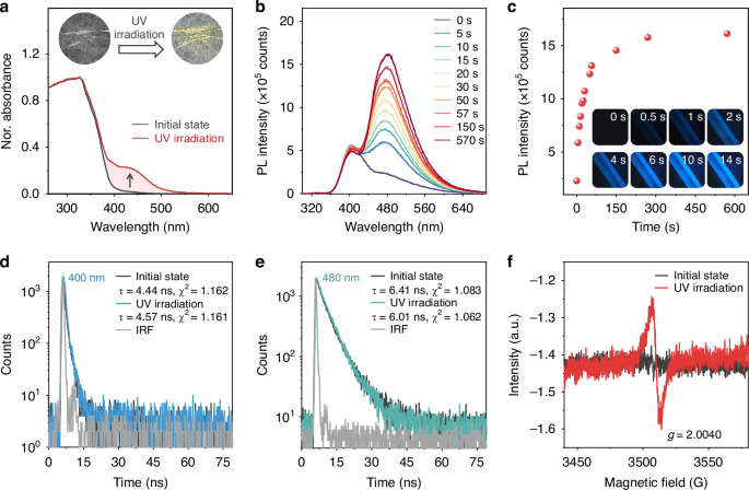
The organic dye NPBr, a derivative of naphthyl benzoate, was prepared by a one-step esterification reaction in a high yield of 76%. The synthesis followed well-established procedures6,34 and is detailed in the Supporting Information. Numerous needle-shaped crystals of NPBr were obtained by recrystallization from a mixture of dichloromethane and methanol at room temperature. The resulting crystals appeared colorless and were nearly non-emissive under UV irradiation. As illustrated in Fig. 2a, the ultraviolet-visible (UV-vis) absorption spectrum of NPBr displayed a broad absorption band in the range of 260–402 nm, consistent with its colorless appearance. Time-dependent density functional theory (TD-DFT) calculations indicated that the absorption peak at 325 nm was attributed to π–π* transitions of naphthyl benzoate (Fig. S1). Upon photoirradiation for several seconds, the colorless crystals gradually transformed to yellow. A new absorption peak emerged at 441 nm, indicating the generation of new emissive species with a narrow optical gap. The pristine NPBr crystals showed a distinct PL peak at 400 nm, accompanied by a weak, broad shoulder band at 480 nm (Fig. 2b). The shoulder arises from the unavoidable light irradiation of NPBr by the excitation source of the spectrometer during measurement. Interestingly, when the NPBr crystalline powders were continuously irradiated with an additional UV lamp at room temperature in air, the PL intensity of the shoulder peak gradually increased. If this experiment were conducted in the dark, a bright blue luminescence would be observed in insets of Fig. 2c. These results demonstrated that NPBr crystals exhibited a photoinduced luminescence behavior. Moreover, the intensity of this photoinduced luminescent peak increased sharply (Fig. 2c) as the irradiative time within 75 s. When the irradiation time was extended from 75 s to 10 min, the rate of increase in PL intensity slowed. After 10 min of photoirradiation, NPBr crystals achieved maximum PL intensity with PLQY of 47.7%, exhibiting a highly competitive PLQY value among reported crystalline luminescent radicals31,32,33,35,36,37. As depicted in Fig. S2, the excitation spectra of photoirradiated NPBr powders correlate well with their absorption spectra, revealing additional long-wavelength absorption features associated with emission processes. Interestingly, the photoinduced luminescence behavior of NPBr was also observed in CH2Cl2 and polymethyl methacrylate (PMMA) films (Fig. S3). As depicted in Fig. 2d, e, the PL lifetime of this dominant peak at 480 nm was measured and calculated to be 6.01 ns, indicative of its fluorescence characteristics38,39,40. Together with the PLQY values, the radiative (kr) and non-radiative (knr) decay rate constants were determined to be 7.9 × 10⁷ s−1 and 8.7 × 10⁷ s−1, respectively. The kr value was relatively high compared to those of other emitting π-radicals. Notably, the new generation of emitting species was highly sensitive to photoirradiation. Thus, during PL measurements, the emissive species could be produced, and the shoulder peak at 480 nm became prominent (Fig. 2b). After 10 min of photoirradiation, the powder XRD spectra of NPBr crystals revealed no discernible changes compared to their pristine state (Fig. S4), indicating that the molecular packing within the crystalline lattice remained unchanged. As illustrated in Fig. S5, this photoinduced luminescence exhibits irreversibility, as it persists after heating or cessation of irradiation.
 The alternative text for this image may have been generated using AI.
a Absorption spectra of NPBr crystalline powders before and after UV irradiation, with insets showing the corresponding photographs of the crystals under ambient light. b PL spectra and c integrated PL intensity at 480 nm of NPBr crystalline powders as a function of UV irradiation time (0–10 min), with insets in (c) showing corresponding PL photographs of a single NPBr crystal. A narrowband filter (380 nm center wavelength, 10 nm bandwidth) and dichroic mirrors were installed in front of the camera lens for imaging. d, e Time-resolved PL decay curves of NPBr (λem = 400 nm and 480 nm) before and after UV irradiation. f EPR spectra of NPBr crystalline powders before and after UV irradiation
Photoinduced luminescence mechanism
UV irradiation is generally known to disrupt molecular structures35,36,37; hence, assessing the structural integrity of the molecules is essential. Figure S6 illustrates the emergence of new 1H NMR signals at δ 12.67, 8.44, 6.81, and 3.85 ppm after 30 min of UV exposure, indicating that the NPBr molecules underwent a photochemical reaction. To elucidate the photochemical reaction pathways, the UV-irradiated NPBr solution was separated and purified by preparative thin-layer chromatography (PTLC), yielding a small amount of isolable product. Its single crystals were subsequently obtained via slow solvent evaporation. The ¹H NMR spectrum of this product (Figs. 3a and S7) displays notable changes in the chemical shifts of aromatic protons compared to those of NPBr. In addition to the original Hb’–Hi’ signals, a new proton resonance, denoted Ha, is observed. Combined with single-crystal XRD analysis (Fig. S8), the formation of CONP after UV irradiation was confirmed. This structural identification provides clear evidence that UV irradiation induces homolytic cleavage of the ester bond in NPBr, generating [CO]• and [NPO]• radicals. These radical species subsequently undergo a photo-Fries rearrangement in solution, ultimately leading to the formation of rearrangement product CONP (Fig. 3b). To further characterize the radical intermediates and quantify their abundance, high-performance liquid chromatography (HPLC) was performed using a Poroshell column. While pristine NPBr showed a retention time of 8.906 min (Fig. 3d), the irradiated sample showed three new peaks at 2.002, 2.131, and 15.196 min (Fig. 3c). These retention times are analogous to those of 4-bromobenzoic acid, 4-methoxy-naphthalen-1-ol, and CONP, respectively (Fig. 3e–g), thus confirming the identity of the radical-derived fragments and corroborating the reaction mechanism inferred from single-crystal XRD analysis. It is noteworthy that the infrared (IR) spectra of NPBr crystals revealed negligible change after UV irradiation for 1 h (Fig. S9), suggesting that photodissociation occurred only in a small fraction of these molecules. This proposal is consistent with quantitative HPLC results. The photophysical properties of NPBr and its photoreaction products were subsequently compared. As shown in Figs. S10a and S11a, 4-bromobenzoic acid and 4-methoxynaphthalen-1-ol displayed the absorption peaks at 270 and 284 nm, respectively, which differ significantly from the absorption peak at 441 nm observed for UV-irradiated NPBr powder. Moreover, 4-bromobenzoic acid emitted phosphorescence with PL peaks at 510 and 548 nm (Fig. S10b–d), whereas 4-methoxynaphthalen-1-ol and CONP showed PL peaks at 386 and 583 nm, respectively (Figs. S11b and S12). These results indicate that the broad shoulder peak at 480 nm in the PL spectrum of irradiated NPBr cannot be attributed to 4-bromobenzoic acid, 4-methoxynaphthalen-1-ol, or CONP. Therefore, the luminescence of photoirradiated NPBr powder is unlikely to originate from protonation or rearrangement products of the radicals. Instead, it is reasonable to attribute the observed photoinduced luminescence to emission from [CO]• or [NPO]• radicals generated under UV irradiation.
 The alternative text for this image may have been generated using AI.
a ¹H NMR spectra of the pristine NPBr (bottom) and the isolated product CONP in CDCl3 (top). b The reaction mechanism for the photo-Fries rearrangement of NPBr in the solution phase upon UV irradiation. HPLC analysis of d pristine NPBr and c its photoirradiated products alongside e 4-bromobenzoic acid, f 4-methoxynaphthalen-1-ol, and g isolated product CONP using a hexane/chloroform (2:3) mobile phase on a Daicel Chiralpak IE column
To obtain direct evidence for the presence of these proposed radical species, we performed EPR spectroscopic studies. As depicted in Fig. 2f, a weak but persistent EPR signal at g = 2.0040 was observed in UV-irradiated NPBr crystals, suggestive of radical formation. The persistence of this signal, even after removal of the irradiation (Fig. S13), aligns with the observed irreversible fluorescence switching, suggesting the generation of stable radical species. To further confirm the identity of the radical species, EPR spin-trapping experiments were conducted using 5,5-dimethyl-1-pyrroline N-oxide (DMPO). No EPR signals were detected prior to irradiation, but a multiline spectrum emerged after irradiation at 300 K (Fig. S14a, b). The observed signals were assigned to DMPO adducts of the benzoyl radical ([CO]•, with hyperfine coupling constants AN = 14.06 G and AH = 15.81 G) and the hydroperoxyl radical ([NPO]•, with AN = 12.86 G and AH = 10.01 G), consistent with reported values for DMPO-trapped species. The experimental spectrum was successfully simulated as a superposition of DMPO/[CO]• and DMPO/[NPO]• adducts (Fig. S14c–e). When the naphthalene moiety of NPBr was replaced by a phenyl group, the resulting compound, benzoate ester BzO, exhibited a broad, less intense emission at 472 nm after UV irradiation (Fig. S15). This PL intensity was significantly lower than that observed for photoirradiated NPBr. Moreover, simulation studies further suggested that the radical signal detected in the DMPO/BzO system in CH2Cl2 solution was indeed a combination of DMPO/[CO]• and DMPO/[BPO]• adducts (Fig. S16). We also synthesized two control molecules, NA and BrO, by replacing the bromine atom and the methoxy group in NPBr with hydrogen atoms, respectively. The crystalline powder of NA exhibited photoinduced luminescence properties and fluorescence lifetime similar to NPBr, while BrO showed no fluorescence at all (Fig. S17). These findings showed that the emerging PL peak at 480 nm observed in photoirradiated NPBr correlated with the [NPO]• radical. The TD-DFT calculations were performed using the M06-2X (6–31 G(d,p)) functional to further investigate the radical emission. The optimized energy gap for the D0 → Dn (n = 2 and 3) transition of the oxygen-centered radical ([NPO]•) has been calculated to be 2.69 eV (461 nm) and 2.78 eV (446 nm), with corresponding oscillator strengths (f) of 0.0220 and 0.0560. These values are remarkably close to the absorption peak observed at 441 nm in photoirradiated NPBr powders (Fig. 2a). However, the benzoyl radical ([CO]•) exhibited the smallest energy gap of 3.3 eV for D0 → Dn (n = 2, 3, and 4) electronic states, and the corresponding transitions were nearly forbidden (f < 0.0003). The results further confirm that the oxygen-centered radical ([NPO]•) is responsible for the photoinduced radical emission. However, the photogenerated radicals are unstable in CH2Cl2 due to the absence of spatial confinement, which is present in both crystalline and rigid PMMA matrices. Experimental evidence supports this conclusion: When NPBr was dissolved in CH2Cl2 at a low concentration (1 mg mL−1), no EPR signal was detected upon photoirradiation at room temperature (Fig. S18a), but it could be detected at 120 K (Fig. S18b). This indicates that the photogenerated radicals are unstable in dilute solution and can only be detected when their decay and recombination processes are kinetically inhibited. Notably, an observable room-temperature EPR signal required increasing the NPBr concentration to 68 mg mL−1 (Fig. S18c). Moreover, the PL spectrum for this concentrated solution exhibits a peak at 517 nm upon photoirradiation, and the fluorescence intensity decreases gradually over time after the photoirradiation is discontinued (Fig. S18d). This time-dependent fluorescence quenching, paralleled by a concurrent decay of the EPR signal (Fig. S18c), consistently indicates the instability and transient nature of the radicals in the solution phase.
Based on comprehensive experimental evidence, we propose a photoactivated self-doping mechanism to account for the photoinduced radical luminescence in benzoate ester. Upon photoirradiation, a small fraction of NPBr molecules undergo a photodissociation process, generating limited benzoyl ([CO]•) and hydroperoxyl ([NPO]•) radicals (approximately 5% molar ratio by HPLC). Photophysical testing and theoretical calculations indicate that the [NPO]• radicals exhibit intense blue fluorescence (λem = 480 nm, PLQY = 47.7%), which dominates the observed photoinduced radical luminescence (Fig. 2b). Crucially, compared to a liquid-phase environment, the NPBr crystalline matrix effectively isolates individual radical species through spatial confinement. This isolation mechanism suppresses radical recombination pathways, enabling remarkable stability of the emissive species over 30 days under ambient conditions (Fig. S5). The irreversible character of the photoinduced luminescence aligns with a self-doping mechanism: (i) Localized photolysis creates isolated [NPO]• radicals embedded within the host NPBr lattice; (ii) These emissive dopants remain spatially fixed due to the restricted molecular mobility in the crystalline state (XRD analysis, Fig. S4), effectively preventing quenching and rearrangement. This photoactivated self-doping process differs fundamentally from conventional physical doping strategies, as it easily achieves spatial control of dopant distribution through the photodissociation process.
Flexibility of crystals
Long crystalline needles were readily obtained, and their photophysical properties were investigated. The typical lengths of the crystals were 10–20 mm with widths ranging from 0.4 to 1 mm. Similar to the crystalline powders, the needle-shaped NPBr crystals exhibited photoinduced luminescence. Notably, these crystals also displayed remarkable elasticity. As depicted in Fig. 4a, applying stress to the ends of a straight crystal using tweezers induced bending along the widest faces, exceeding 180° without cracking or breaking. Upon the release of the applied stress, the crystal instantaneously reverted to its original, straight configuration (Fig. S19 and Video S1). This bending-relaxation cycle could be repeated several times without apparent degradation, demonstrating the remarkable elastic resilience of NPBr crystals. In addition, the corresponding luminescent behavior remained unchanged upon mechanical deformation. To quantitatively assess the mechanical properties of the flexible NPBr crystals, nanoindentation measurements were conducted (Fig. S20). The average Young’s modulus of NPBr crystals was determined to be approximately 9.76 GPa, which was very close to that of the crystals after photoirradiation (9.50 GPa). This suggests that photoirradiation does not significantly alter the mechanical properties of the crystals. Consistent with these findings, UV-irradiated NPBr crystals exhibited good flexibility and could be readily bent under external force, as depicted in Fig. S21.
 The alternative text for this image may have been generated using AI.
a Elastic bending process of a needle-shaped NPBr crystal compressed by tweezers. b Molecular conformation of the NPBr crystal. c The overlapping area and vertical distance of the π−π interaction along one-dimensional molecular chain. d Crystal packing diagrams viewed along the a- and c-axes directions, revealing the hydrogen-bonded network that constructs the (100) and (001) crystallographic planes. e Schematic representation of crystal deformation mechanisms: (top) pristine single crystal and (bottom) bent morphology. The arrows indicate the expansion and compression directions of outer and inner arcs during the bending process
To elucidate the flexible nature of NPBr, its crystal structure was determined using single-crystal XRD analysis. The crystal belongs to the monoclinic space group P21/n (Z = 4, ρ = 1.635 cm³). The asymmetric unit comprised one NPBr molecule, with no solvent molecules present within the crystal lattice. Each NPBr molecule exhibited a bent conformation, evidenced by a dihedral angle of approximately 90° between the phenyl ring and the naphthalene unit (Fig. 4b). And the neighboring molecules revealed a distinct overlap between naphthalene planes with a short distance of approximately 3.36 Å (Fig. 4c). The neighboring π-π stacking had significant interaction energy (−20.5 kJ mol−1), driving molecular chain formation along the b-axis. As depicted in Figs. 4d and S22, these molecules were connected along the c-axis direction by two types of CH•••O interactions with distances of 2.59 and 3.73 Å, which produce a structurally stable molecular layer. Moreover, these stacked columns were linked by intermolecular hydrogen bonds CH•••O and Br•••O halogen bond with the distance of 2.97 Å and 3.53 Å along the [010] direction, generating the bendable (001) planes. Consequently, in addition to the infinite π-stacking, the C–H•••O hydrogen bonds (−17.7 kJ mol−1) and Br•••O halogen interactions (−5.1 kJ mol−1) between molecular chains suppressed the breaking tendency of molecular chains towards expansion, thus benefiting the high elasticity of OSCs.
To verify the critical role of Br•••O interactions, we performed crystal structure analysis on NA and BrO. Both NA and BrO crystallized as block-shaped crystals, sharply contrasting with the needle-like morphology of NPBr (Fig. S24). Single-crystal analyses revealed the absence of Br•••O interactions in these derivatives, accompanied by a complete disruption of the directional molecular chains observed in NPBr. The packing arrangements of NA and BrO exhibited disordered stacking patterns with increased intermolecular distances (Fig. S25), confirming that halogen bonding is essential for maintaining the one-dimensional (1D) chain architecture. These comparative results conclusively establish that Br•••O interactions serve as key structural determinants for both crystal morphology and elastic deformability in this system6. Why do NPBr crystals exhibit controllable elasticity and bending? As illustrated in Fig. 4e, the elastic bending of the crystal involved the respective expansion and contraction of the outer and inner arcs. As molecular chains aligned along the b-axis, the π-π interactions within these chains dynamically adjusted to the outer arc’s expansion by increasing the π–π distance, while responding to the inner arc’s contraction by decreasing the π–π distance. In the crystal structure of the (010) plane, the oxygen atoms form type-II O/Br contacts at a distance of 3.53 Å and exhibit C–H/O interactions at distances of 2.97 Å (Fig. S23). The multiple Br–O and C–H/O interactions, which are arranged in diverse directions and balanced with each other, are relatively stable and thus could avoid changing the position of the molecule, i.e., preventing displacement of the molecular layer structure, during the process of elastic bending. Consequently, these interactions play a critical role in enabling crystal elasticity41,42,43.
Optical waveguiding properties
Interestingly, the crystal exhibits optical waveguiding properties, which were observed from the brighter emission at the tips compared to the body when the straight crystal was illuminated with a UV lamp. Further investigations revealed that this waveguiding behavior is not limited to the straight crystal but is also present in highly bent crystals. To quantitatively evaluate the effects of elastic bending on waveguiding performance, the optical loss coefficients (OLCs) of a single NPBr crystal were investigated in both its straight and highly bent configurations, as illustrated in Fig. 5a. By exposing various positions along the crystal to a consistent excitation source of a 355 nm laser (Fig. 5b, e) and subsequently collecting the emission spectra at one tip of the crystal, distance-dependent PL profiles were established as depicted in Fig. 5c, f. The PL intensity at the tip diminished progressively as the distance between the tip and the irradiated position increased, attributed to a greater loss of emitted light over longer propagation distances. Additionally, the PL spectra exhibited a slight redshift with increased propagation distance, caused by enhanced reabsorption within the spectral overlap region between PL and absorption bands (Fig. S26). Utilizing fitting procedures described in the literature for the data presented in Fig. 5d, g44, the OLCs at 488 nm were estimated to be 0.584 dB mm−1 for the straight state and 0.806 dB mm−1 for the bent state. These low OLC values surpass those of many reported organic crystals and polymeric systems45,46,47,48,49, indicating a favorable waveguiding performance in both configurations. Notably, the OLC for the bent state is slightly increased relative to that of the straight state. The maintenance of a low OLC in the highly bent condition is particularly intriguing, as optical losses in waveguides are typically exacerbated in bent configurations.
 The alternative text for this image may have been generated using AI.
a Schematic diagram of the optical waveguide test setup. b, e Straight and bent crystals under excitation from a UV lamp (top image) and a 355 nm laser focused on different positions. c, f PL spectra collected at the tip of a single crystal in the straight state and bent state with the different distances between the tip and the excitation site of the laser. d, g The Itip/Ibody decay curves of a single crystal (length = 6 mm) in the straight and bent states. The optical loss coefficients (α) were determined using a single exponential fit to the equation Itip/Ibody = Aexp(−αD), where Itip and Ibody represent the PL intensities of out-coupled and incident light, respectively