
For over a century, generations of neuroscientists have dedicated countless hours to deciphering how the brain works. To understand the central nervous system, many believed that a full description of its anatomical connections was necessary—a daunting task considering that even a simple organism like the fruit fly Drosophila melanogaster has approximately 100,000 neurons.
To reconstruct the neuronal wiring of the brain, some scientists realized they needed to find an organism with three important qualities. It needed to have a simple enough central nervous system. It also had to be easy to genetically manipulate and perform behaviors that could be easily quantified and linked to the function of neurons and their connected networks.
In the mid-1960s, in his laboratory at the Medical Research Council Laboratory of Molecular Biology (MRC-LMB), biologist Sydney Brenner thought that nematodes, particularly the transparent Caenorhabditis elegans, could help achieve the complete mapping of a nervous system. The one-millimeter-long worm checked many of Brenner’s boxes: It had a small number of neurons, about 300, and it was easy to rear in the lab and observe under a microscope.1 Brenner was also familiar with the genetics of the organism as he was studying its mutants and their phenotypes.2
Forty years ago, Brenner led a small troop of daring scientists who believed in his idea and set out to chart C. elegans’ neuronal connections in full. The herculean task resulted in the first neuronal connectome—decades before the term connectome was independently coined by researchers Olaf Sporns and Patric Hagmann in 2005.3,4 Above all, it provided researchers in the C. elegans field and beyond a foundation to understand the functions of the worm’s nervous system and of more complex brains.
Making the First C. elegans Neuronal Wiring Map
To reconstruct the C. elegans nervous system, Brenner needed a team with the right skill set. To study the anatomy of an organ, researchers often cut sequential sections of a preserved specimen. Brenner and his team planned to use this approach paired with electron microscopy to obtain detailed images of the C. elegans neurons and their neuronal partners.
So, he hired Nichol Thomson, a skilled electron microscopist, to be in charge of this task.2 “[Thomson] could cut many series of sections without losing them. This was tremendously important for what we needed to do,” said John White, an emeritus professor at the University of Wisconsin-Madison who was part of Brenner’s team behind creating the first C. elegans connectome.
White joined Brenner’s lab in 1969 almost by accident. “I went there because I’d heard that [Brenner] was setting up this project and trying to reconstruct a nervous system,” he said. “My own background in physics and electronics made me very interested in circuit diagrams and how things work, and I knew enough about animals to realize that their brains were essentially very complicated circuits,” White added. “The whole project intrigued me.”

As a member of Brenner’s lab, John White was in charge of developing a computing system to help process the large amounts of data the complete description of the C. elegans neuronal wiring diagram produced.
Wormbook, CC By 2.5 license.
As part of the team, White’s main task was to develop a computer system to help process the large amounts of data the project was going to generate. Computers in the late 1960s and early 1970s were not like the machines of today, noted White, adding that he initially focused on developing basic computer programming that allowed the computer to read information and show things on the screen. While limitations of speed and data storage capacity soon revealed that White’s computer system could not support data of such complexity and quantity, the researchers did use the system to check the worm’s synapse distribution as the project went on.5
The last core member of the team was technician Eileen Southgate, who created electron micrographs from Thomson’s sections, printed them as large images, and then, along with White, worked on putting together these images by hand. “She was very meticulous and just liked putting things together,” White recalled. “It turned out that her hobby was jigsaw puzzles. She used to do all this all day with the electron microscope pictures and then go home and do a thousand-piece jigsaw puzzle.”
To start the reconstruction of the C. elegans neuronal map, White and Southgate used erasable fine-tipped colored pens to mark the electron micrographs, tracking neurons and their processes across the stack of images and labeling the cells with numbers to differentiate one from the other. If two colored tracks fused, the researchers would erase one of them and replace it with the other. After a neuron was completely tracked, they identified the synapses and gap junctions and tried to sort out any ambiguities that might have arisen during the process.1,5
While the team’s goal was to create a full nervous system wiring diagram, they first mapped the sensory nervous system located on the worm’s head and its ventral nerve cord, a structure essential for the animal’s sine wave-like undulation and back-and-forth locomotion.6,7 With these initial reconstructions, White recalled that the team got motivated to carry on the main project as they could see different classes of neurons with unique features emerging from their work. It took the team more than a decade to complete the reconstruction of the C. elegans anatomical connectome, identifying around 5,000 chemical synapses, 2,000 neuromuscular junctions, and 600 gap junctions.3 When the work was accepted for publication in 1986, it filled an entire volume of the Philosophical Transactions of the Royal Society of London journal and, to the authors’ amusement, kept the phrase ‘The mind of a worm,’ which they had used to identify the notebooks containing texts and figures sent for publication, as the running title.3,5
Mapping the Male C. elegans Circuit
The work by White, Brenner, and their colleagues created an anatomical map of individual neurons and their connections as had never been seen before. For scientists like Oliver Hobert, a molecular biologist at Columbia University, the work was a valuable source of information to better understand the functions of the brain. “It’s an amazing resource. The first day I started working on C. elegans, I got the hard copy of that paper, and I’m still using that hard copy,” he said. “Some of us call it the ‘bible of the worm.’” Today Hobert uses the nematode as a model organism to study how genes determine the unique features of cells in the nervous system.

Technician Eileen Southgate was a key member of the team who looked at the ‘mind of a worm’ and brought the first connectome to life.
Wormbook, CC By 2.5 license.
The first complete C. elegans connectivity map described the nervous system of the hermaphrodite, the self-fertile individual, which is considered the female of the roundworm. In the 1980s, researchers at the MRC-LMB then began mapping the neural circuits found in the male nematode, specifically in its tail, which controls mating and differs in look and function from the hermaphrodite tail.8 But, due to its higher complexity, the team only made a partial reconstruction.
In the early 2000s, Scott Emmons, now an emeritus professor at the Albert Einstein College of Medicine, wondered if it would be possible to complete the male’s tail connectome. “It’s more complex in the male tail than elsewhere in the nervous system,” Emmons explained. “They didn’t have Excel. They couldn’t write down the data in a table. [It was] an impossible task. But John White thought, by 1999, that it could be done. So, I started.”
Emmons, who was well acquainted with the worm because of his early work on the nematode’s genome and developmental biology, set up a collaboration with Albert Einstein College of Medicine electron microscopist David Hall, who had helped reconstruct the posterior nervous system of the hermaphrodite and knew how to interpret the serial sections of electron micrographs.
The duo got the original images of the MRC-LMB’s first male tail work and, after scanning them, began completing the neuronal wiring diagram. They uncovered that the circuit works in local feedback loops, where sensory neurons are only one synapse away from either a muscle or the gonad.9 The compact and highly connected layout, Emmons and his colleagues argued, may help explain how the system quickly selects and sets in motion mating behavior. “I was very disappointed,” said Emmons. “I thought, ‘Well, this is not decision-making, this is just a reflex. Neuroscience is not going to be interested in this. Then it occurred to me, ‘Wait a minute. That’s the discovery. What looks like decision-making is nothing but a reflex arc.’”

The first neuronal connectome in the worm C. elegans occupied the entire volume of the 1986 Philosophical Transactions of the Royal Society of London journal and is still used today by researchers, who often refer to it as ‘The bible of the worm.’
Oliver Hobert
Inspired by Emmons’ work on the male tail circuit, Hobert began studying the C. elegans connectome, and the two collaborated to map the entire neuronal wiring network of the male worm and compare it to that of the hermaphrodite.10 “There was never any comparison between the sexes of a connectome, so that was something of interest,” said Emmons.
To be able to compare both sexes, the researchers reanalyzed the original 1980s images from the hermaphrodite using software that they developed. Beyond providing a neuronal-level comparison between the male and hermaphrodite, the researchers also inferred the strength of the connections by taking the size of the synapses into account as a measurement of how strong these connections were. This revealed that up to 30 percent of them differ in strength between the sexes.10
Beyond Synapses to the ‘Wireless Connectome’
Although these neuronal wiring maps are stepping stones toward understanding an organism’s nervous system, they do not fully explain how it works. “I consider a synaptic connectome like sequencing a genome, but you only sequence the Gs,” said Hobert. “Very useful, but it’s not gonna tell you where the genes are [or] give you the genetic code. It’s a very, very incomplete picture for predicting information flow in the nervous system.”
Overcoming some of these limitations to better understand the nervous system was a goal of William Schafer, a neuroscientist at the MRC-LMB. Schafer became interested in studying the brain when he was considering applying for a postdoctoral position. His background in molecular biology and genetics led him to choose C. elegans as a model organism as it was simple enough that a non-neuroscientist like him “could get their head around” it, he said.
As a postdoctoral researcher in the laboratory of Cynthia Kenyon, now an emeritus professor at the University of California, San Francisco, he realized that neuromodulators did not have to follow the synaptic connectivity established by the physical interactions between neurons as they may act on cells that are not directly connected. “For a lot of people in the C. elegans field, this sort of provoked this realization of the importance of diffusible signals in the brain,” he said.
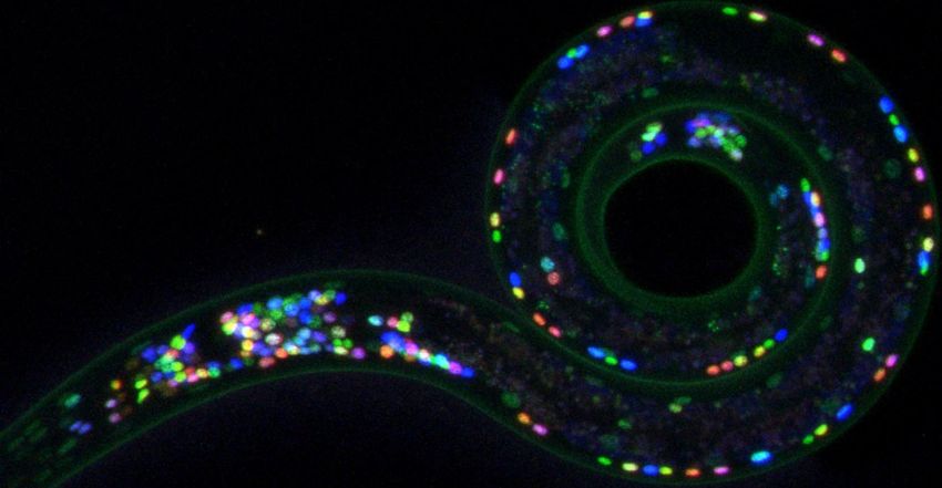
By combining a technique developed by Hobert’s team, which enables the identifications of all neurons, with other methods, Schafer and colleagues described the multi-layer nature of the C. elegans connectome.
Ev Yemini and Oliver Hobert
In his lab, Schafer and his team started to tease out these extrasynaptic mechanisms by looking at monoamines and neuropeptides, which are known to act as diffusible messengers and modulate nervous system functions.11 Using published and in-house generated data on the expression of monoamines and neuropeptides receptors, enzymes, and vesicle transporter genes, Schafer’s team produced a draft of a map of extrasynaptic monoaminergic and neuropeptide networks in C. elegans.12 “We had synaptic networks, but then we have these wireless neuropeptide and monoamine networks that [are] paralleling the synaptic networks,” Schafer explained.
After their initial publication, more data on neuropeptides and their receptors became available, allowing Schafer’s team, in collaboration with Hobert and other researchers, to complete the neuropeptidergic connectome seven years later.13 “We call it a ‘wireless connectome’ because it doesn’t have physical connections,” Hobert explained. “And [it] shows very little, if any, correlation to the synaptic wiring diagram,” suggesting that the way information flows in the C. elegans nervous system is not determined by its physical road map of neuronal connections but is influenced by wireless neuromodulatory signals.
The Tiny and Mighty Worm Is a Connectomics Pioneer
While C. elegans researchers have advanced the study of the worm’s connectomics by exploring its multiple layers and how they interact to modulate the nematode’s nervous system, similar initiatives to map the brains of other organisms are still in their infancy.
Over the last five years, large collaborative scientific efforts have set out to describe the neuronal connectivity of one cubic millimeter of a mouse visual cortex and, more recently, finished the most comprehensive neuronal wiring diagram of the male fruit fly central nervous system.14,15 “That’s really the first connectome paper on fruit flies that goes as far as John White’s paper in 1986,” said Emmons. “It took 40 years to do the next animal, and it’s not absolutely complete.” To Emmons, the fruit fly work also confirms the observations he and his collaborators made over a decade ago. “The conclusion that they make about the general organization of the circuits is exactly the same [as we] discovered in the C. elegans male tail: The behavior is controlled very locally by local sensory [neurons], directly connected to local effectors in feedback loops,” he said. “That’s really quite interesting.”
As much of a pioneer as C. elegans was in the connectomics field, the worm, through the researchers dedicated to studying it, has not stopped pushing the boundaries of what scientists can learn about its nervous system. For instance, over the past decade, Hobert’s team has uncovered that many of the worm’s sexual differences in the neuronal connectivity are genetically programmed, though environmental factors, such as starvation, can also shape the sexual specificity of these neuronal circuits.16,17
Using the neuronal connectome to study how brains evolve is another area in which the nematode is a trailblazer. By comparing the neuronal connectivity of C. elegans to that of a distantly related predatory nematode, Hobert’s team discovered that, although the two connectomes are highly similar in terms of cellular composition, changes occurred in how these neurons connect and in the location of homologous neurons.18 “We’re looking now at much more distant nematode species to see sort of the next steps of evolution,” Hobert said. “As you become more and more [distant], we’re expecting to see more and more dramatic differences.”
More than 40 years have passed since Brenner first proposed using C. elegans as a model to understand the central nervous system, an idea that, as White recalled, received a lot of criticism at the time. “’Why should we devote our time to this silly little worm, which clearly has a lot of differences to the main nervous systems?’ [As] time went on, it was clear that there are lots of extraordinary similarities,” said White, pointing out how the worm uses the same mechanisms and proteins for synapses and gap junctions as other organisms do.
“One feels that you are building on the basis of something that we found out quite a lot about and can use that knowledge to basically tackle things that are far more complex,” he added. “What is satisfying about it was to see that it was relevant.”