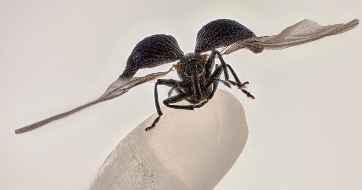
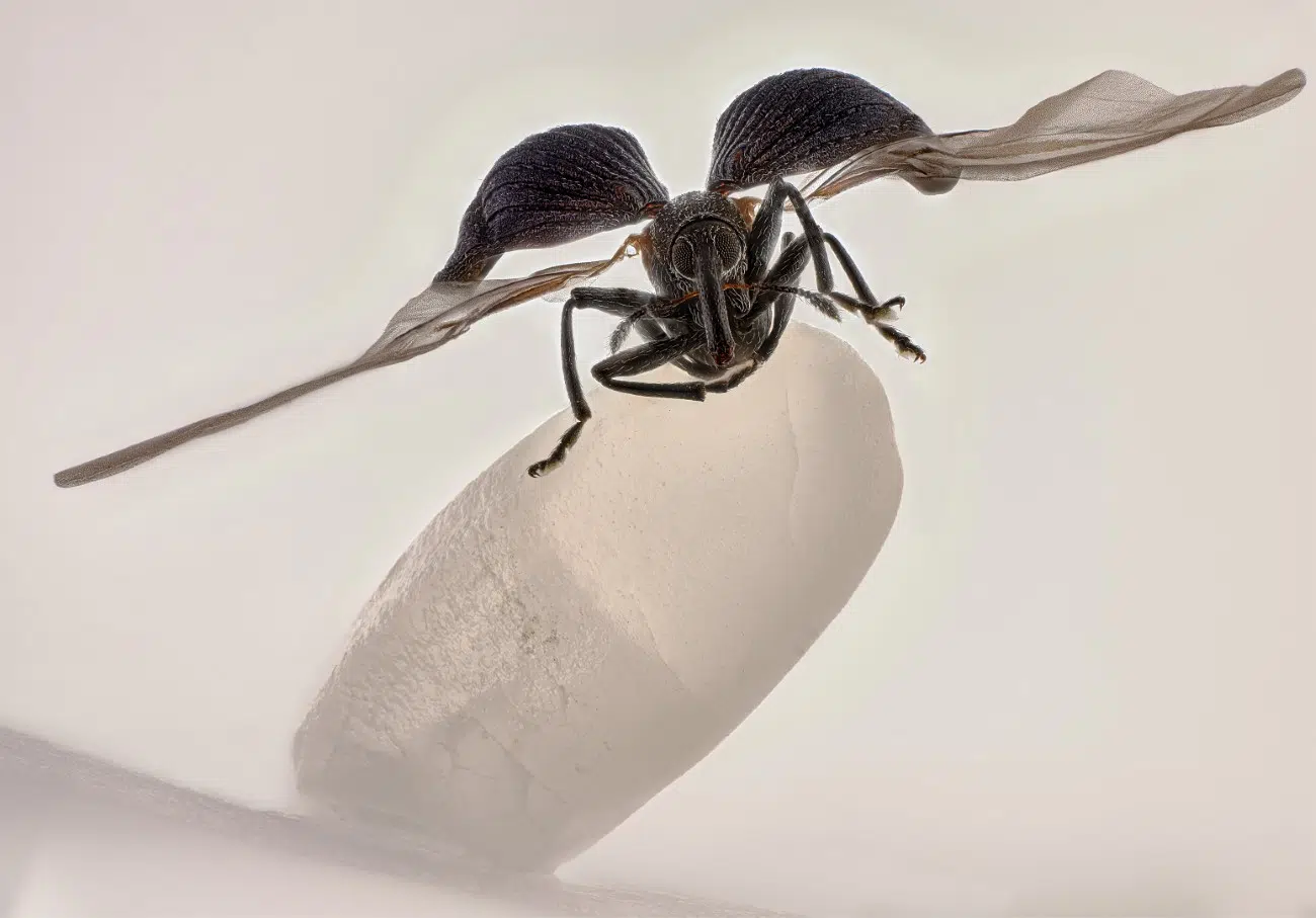
Zhang You
Kunming, Yunnan, China, 1st Place, Rice weevil (Sitophilus oryzae) on a grain of rice, Image Stacking, 5X (Objective Lens Magnification)
It may look like an alien lifeform perched on a crystal, but Zhang You’s winning photograph from the Nikon Small World competition isn’t as sinister as it appears. In reality, the insect lover has captured a striking portrait of a rice weevil sitting on a grain of rice. It’s incredible imagery like this that makes Nikon Small World so unique in the world of photography competitions.
Launched in 1974, the contest shines a light on the world of scientific microscopy and digital imaging. By bringing these images to a wider audience, they help the public understand just how artistic microscope imaging can be. Zhang, for instance, has spent years perfecting his craft by examining insect behavior and his winning photograph shows the benefits he’s gained from that practice.
“It pays to dive deep into entomology: understanding insects’ behaviors and mastering lighting,” he says. “A standout work blends artistry with scientific rigor, capturing the very essence, energy, and spirit of these creatures.”
Interestingly, this was Zhang’s first time entering the competition and he not only won, but also had another image place in the top 20.
“His achievement highlights the spirit of Nikon Small World: inspiring wonder, making scientific understanding accessible to all, and celebrating the artistry of the microscopic realm,” shares Eric Flem, senior manager, communications and CRM at Nikon Instruments.
Other top images include everything from pollen trapped in a spider web to heart cell muscles, demonstrating the variety of what can be photographed under the microscope.
The reveal of these winners comes on the heels of the Nikon Small World in Motion competition, which awards excellence in moving images taken with a microscope. Combined, both provide a revealing look at the invisible world that surround us.
Here are the top 12 photographs from the 2025 Nikon Small World photo competition.
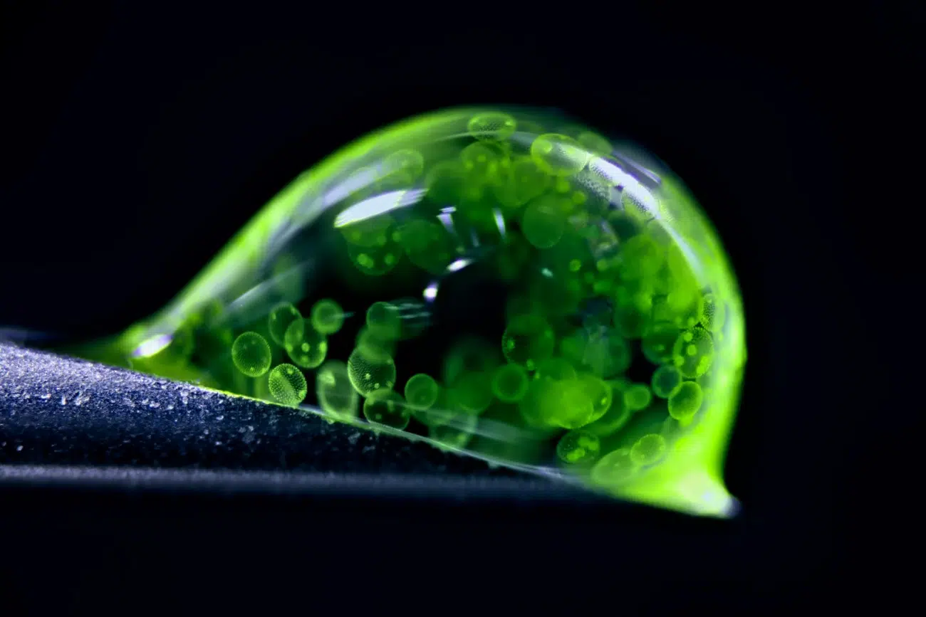
Dr. Jan Rosenboom
Rostock, Mecklenburg Vorpommern, Germany, 2nd Place, Colonial algae (Volvox) spheres in a drop of water, Reflected Light, 5X (Objective Lens Magnification)
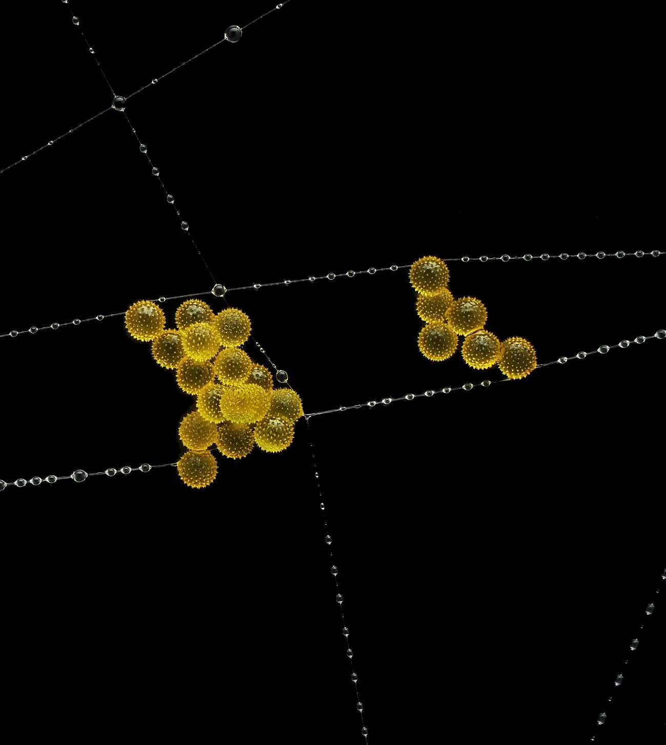
John-Oliver Dum
Medienbunker Produktion, Bendorf, Rheinland Pfalz, Germany, 3rd Place , Pollen in a garden spider web, Image Stacking, 20X (Objective Lens Magnification)
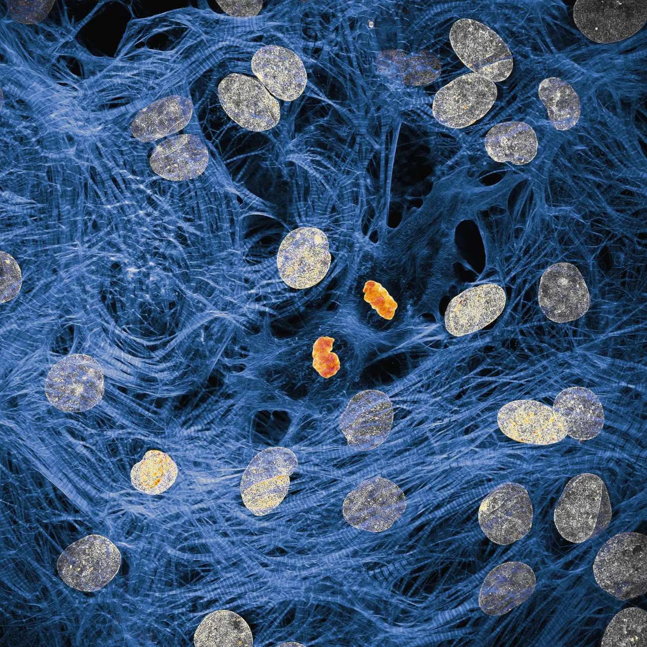
Dr. James Hayes, Vanderbilt University, Department of Cell and Developmental Biology, Nashville, Tennessee, USA, 4th Place, Heart muscle cells with chromosomes condensed following cell division, Confocal, 100X (Objective Lens Magnification)
Dr. Igor Siwanowicz, Howard Hughes Medical Institute (HHMI), Janelia Research Campus, Ashburn, Virginia, USA, 5th Place, Spores (blue/purple structures) of a small tropical fern (Ceratopteris richardii), Confocal, 25X (Objective Lens Magnification)
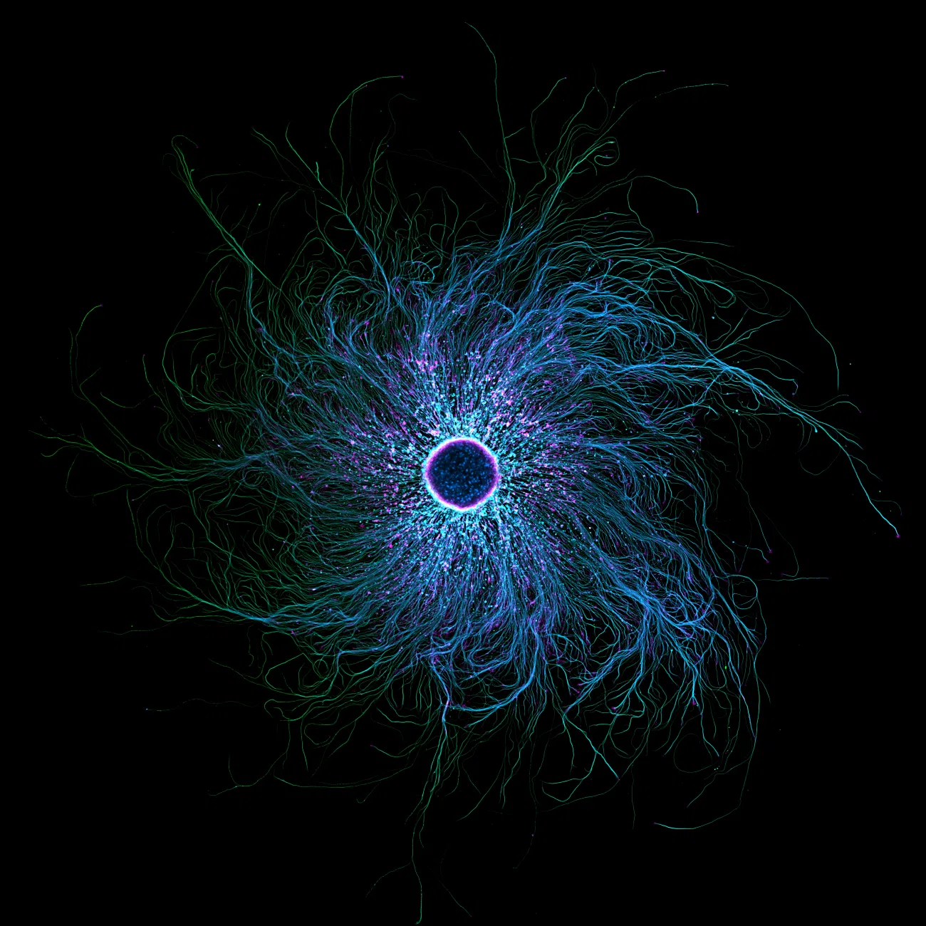
Stella Whittaker, National Institutes of Health, National Institute of Neurological Disorders and Stroke
Bethesda, Maryland, USA, 7th Place, iPSC-derived sensory neurons labeled to show tubulin and actin, Confocal, Fluorescence, Image Stacking, 10X (Objective Lens Magnification)
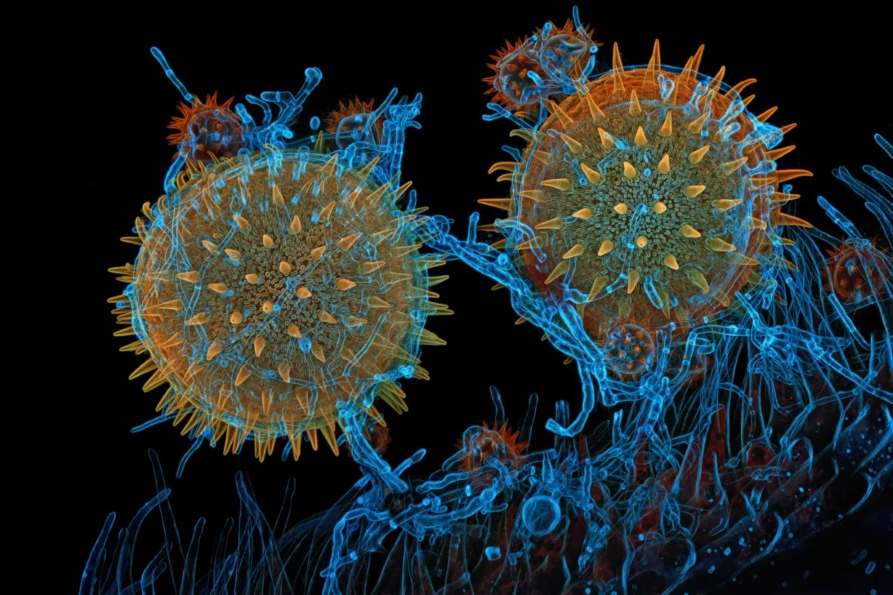
Dr. Igor Siwanowicz, Howard Hughes Medical Institute (HHMI), Janelia Research Campus
Ashburn, Virginia, USA, 8th Place, Mallow pollen germinating on stigma while being parasitized by a filamentous fungus, Confocal, 40X (Objective Lens Magnification)
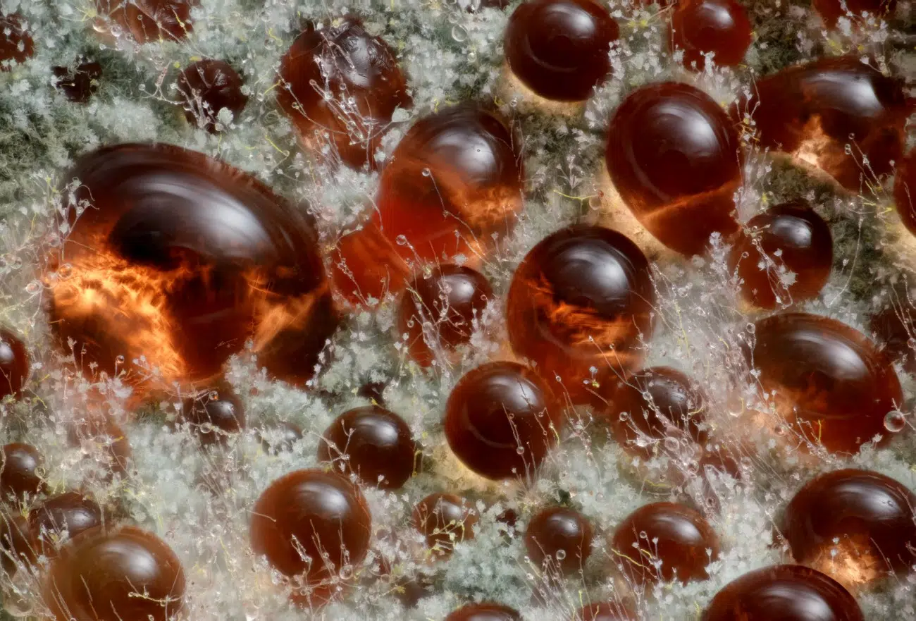
Wim van Egmond, Micropolitan Museum, Berkel en Rodenrijs, Zuid Holland, Netherlands, 9th Place, A fungus (Talaromyces purpureogenus) known for its red, diffused pigment, Image Stacking, 10X (Objective Lens Magnification)
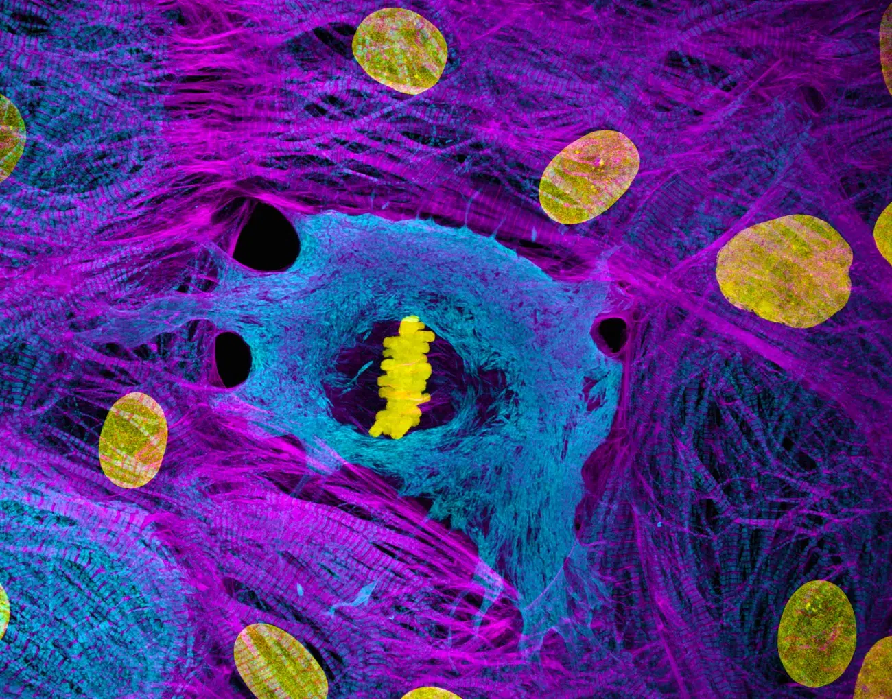
Dr. Dylan Burnette & Dr. James Hayes, Vanderbilt University School of Medicine
Department of Cell and Developmental Biology, Nashville, Tennessee, USA, 10th Place, Heart muscle cells (iPSC-derived) showing condensed chromosomes in metaphase, Structured Illumination, Microscopy (SIM), 60X (Objective Lens Magnification)
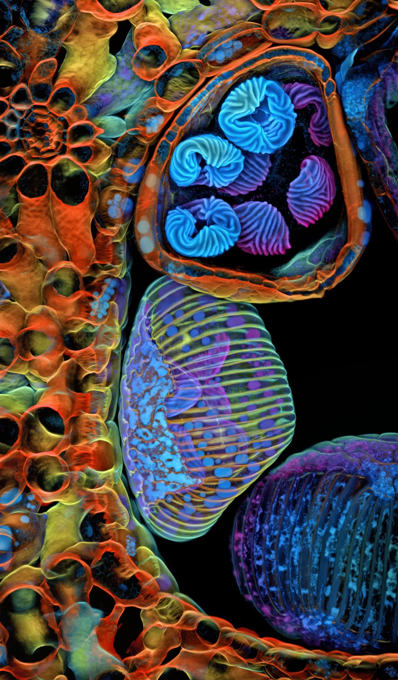
Dr. Francisco Lázaro-Diéguez, Albert Einstein College of Medicine, Bronx, New York, USA, 6th Place, Rat liver cells, Confocal, 63X (Objective Lens Magnification)
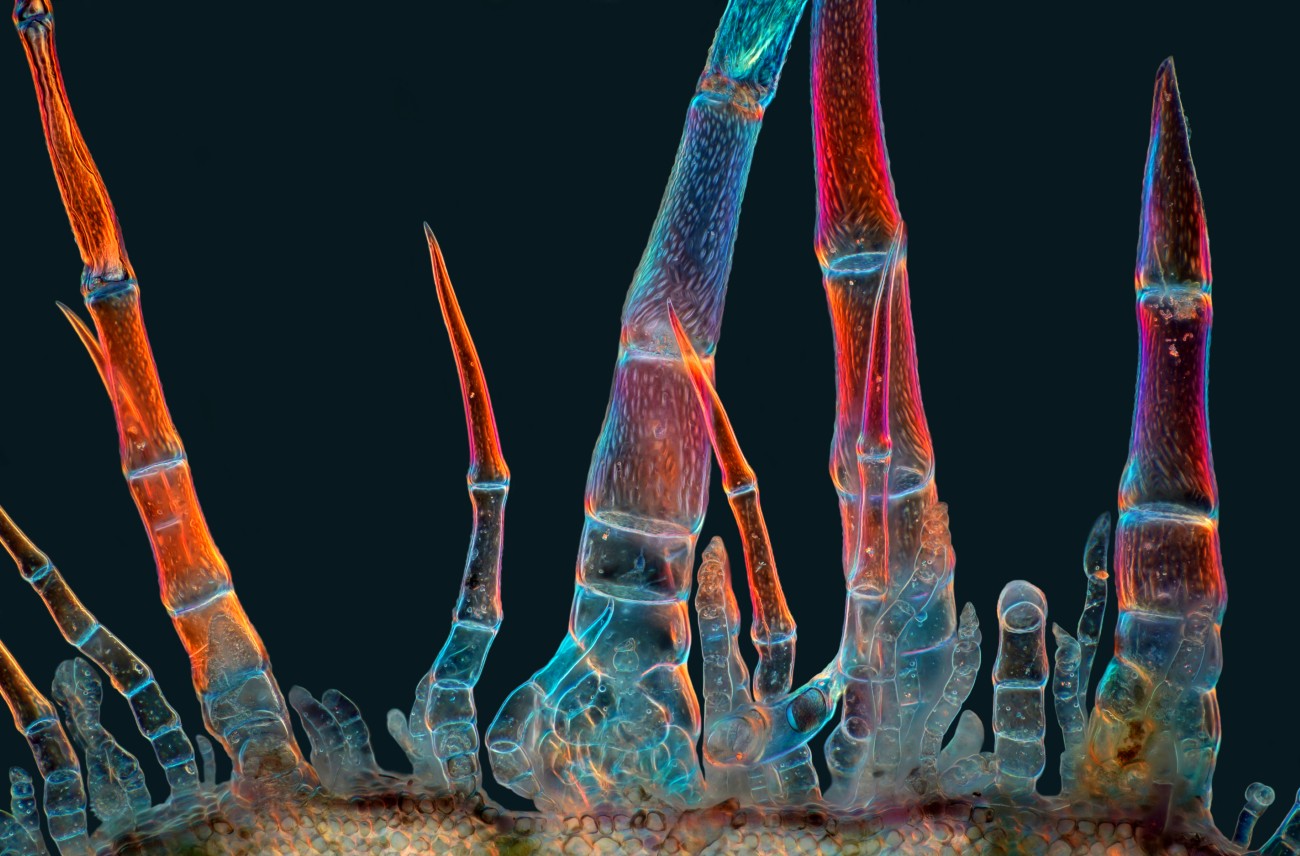
Marek Miś, Marek Miś Photography, Suwalki, Podlaskie, Poland, 11th Place, Sunflower trichomes (hair-like plant outgrowths), Polarized Light, 10X (Objective Lens Magnification)
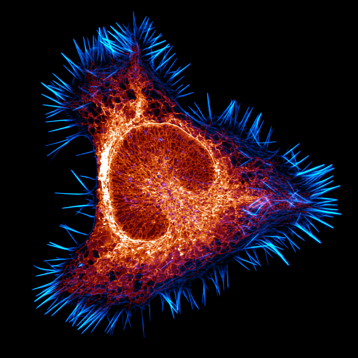
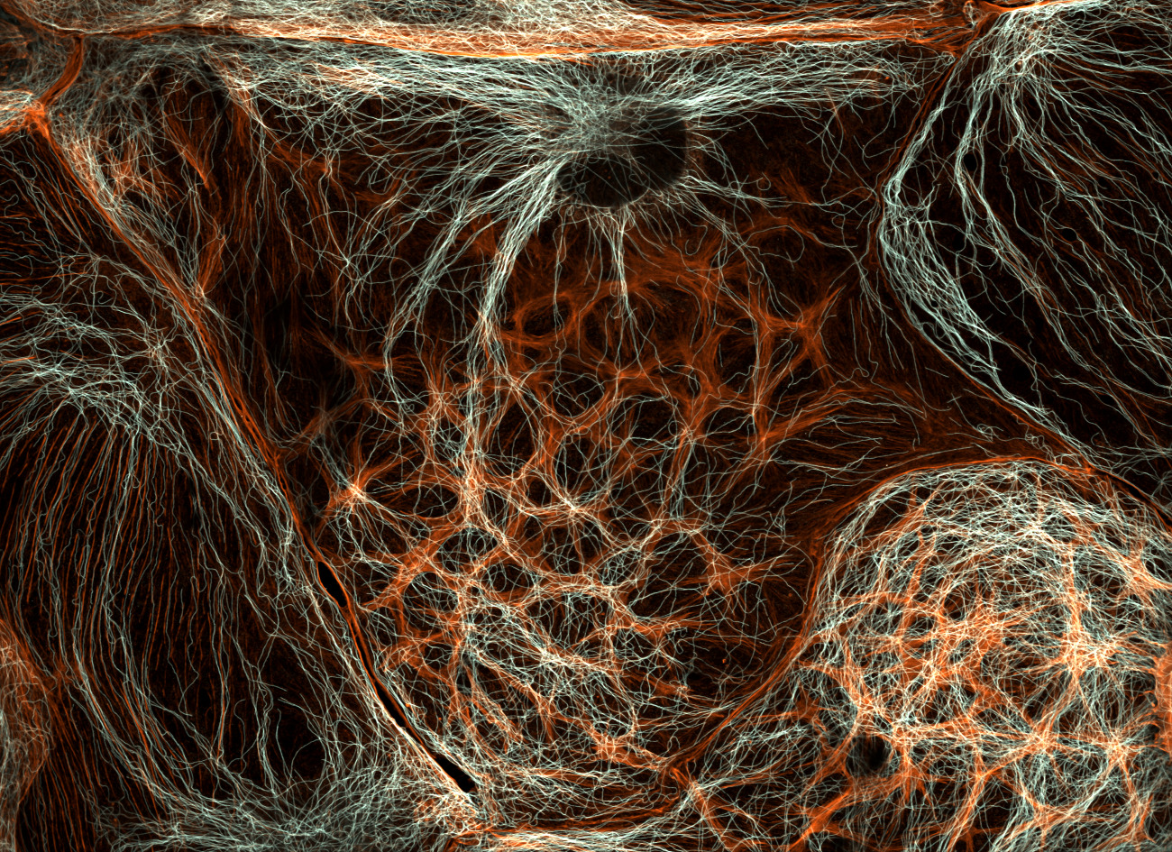
Halli Lindamood & Eric Vitriol, Augusta University, Department of Neuroscience and Regenerative Medicine
Augusta, Georgia, USA, 12th Place, The actin cytoskeleton (cyan) and endoplasmic reticulum (red) of a mouse brain cancer cell, Confocal, Deconvolution, 100X (Objective Lens Magnification)
Nikon Small World: Website | Facebook | Instagram
My Modern Met granted permission to feature photos by Nikon Small World.
Related Articles:
Wildlife Photography Contest Honors the Resilience of Nature
Rare Brown Hyena Photo Wins 2025 Wildlife Photographer of the Year Competition
Colorful “Ladybugs of the Sea” Win 2025 Ocean Photographer of the Year Contest
Bird Soaring Below a Total Solar Eclipse Wins 2025 Bird Photographer of the Year