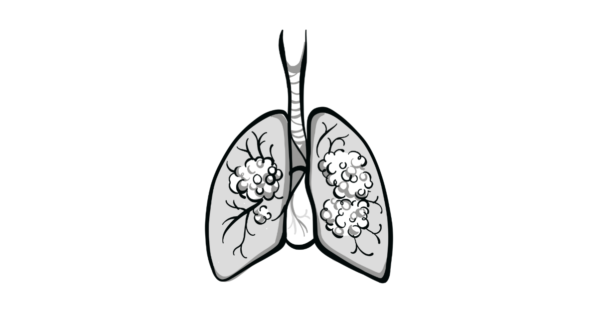

In the modern era of lung cancer diagnosis and treatment, tissue biopsy samples that were once needed for disease staging have become vital for biomarker testing as well. As endobronchial ultrasound (EBUS)-guided biopsy has largely replaced percutaneous approaches for lung cancer, research has not directly addressed the comparison of these 2 approaches and the question of sample adequacy for biomarker testing. Recently, a multidisciplinary expert panel from the American Association of Bronchology and Interventional Pulmonology (AABIP) and the Early Detection and Screening Committee of the International Association for the Study of Lung Cancer (IASLC) published a clinical practice guideline in the Journal of Thoracic Oncology based on a comprehensive review of available data.1
Abhinav Agrawal, MD, system director of interventional pulmonology at Northwell Health in New York, and one of the lead authors of the report, discussed their findings in an interview with Targeted Oncology. He covered the questions of safety, sample adequacy, and the urgency of getting rapid biomarker testing.
Targeted Oncology: What made you investigate these questions about bronchoscopic biopsies?
Abhinav Agrawal, MD: Advances in lung cancer have come in the form of newer targeted therapy options for our patients. As we have increased lung cancer screening, one of the questions that has come to us is how do we diagnose these patients early? Also, when we diagnose these patients with either local or locally advanced lung cancer, or even with metastatic disease, how do we obtain tissue not just for diagnosis, but also for adequate biomarker testing, and do that in the safest manner? The field of interventional pulmonology has rapidly advanced in the last 10 years along with the treatment options, allowing for us to reach a diagnosis faster and get as much tissue as possible.
In light of that, we have seen robotic bronchoscopy or navigational bronchoscopy techniques which came to the market in 2018 and have rapidly seen advancement and adoption across the country. What we wanted to find out was, as robotic bronchoscopy allowed us to both get adequate tissue, not just from the diagnosis standpoint, but also from the safety standpoint, and make sure that we get enough tissue for molecular testing.
We recently published the VERITAS trial [NCT04250194] in the field of interventional pulmonology in the New England Journal of Medicine, and what that showed us was that the diagnostic yield of robotic bronchoscopy is equal to that of CT-guided biopsies.2 That told us about the diagnostic yield and…about the safety. The one question it did not answer was, what is the diagnostic yield for molecular testing? In this era, it’s hard to design a personalized treatment option for patients without that. That was why, through the IASLC as well as the AABIP, we created this focused work group to try to answer this question.
What were the most important findings of the report?
We have 2 subgroups of questions in this paper. One is, when we have a peripheral pulmonary nodule, how should we approach these patients? What are the options available, and what is the safety profile as well as the yield for molecular testing? We assessed guided bronchoscopy to biopsy peripheral pulmonary nodules. Is it as safe or safer than the current standard approach, which is CT-guided biopsy? The second question was, when we do these biopsies, is the tissue that we are obtaining enough to inform decision-making and not subject the patient to additional biopsies to obtain molecular tissue? What we found out was that robotic navigational bronchoscopy, because we are not transgressing the pleura, is safer because the rate of pneumothorax is lower, which was also corroborated by the VERITAS trial.1,2
But the unanswered question…which we were able to answer was that the diagnostic yield and the amount of tissue we are able to obtain. Combining robotic bronchoscopy with forceps or cryobiopsies…can both inform what the diagnosis is, but also give us enough information in terms of biomarker testing, which will allow for the personalized treatment plan for our patients.
The second question is more of a traditional question. When we are doing mediastinal or hilar lymph node biopsy using endobronchial ultrasound, the traditional way about 20 years ago was mediastinoscopy, when EBUS was not available. The community has seen a shift to doing EBUS- guided lymph node biopsy given the high sensitivity as well as the high positive predictive value and negative predictive value. What we tried to answer was to compare that with mediastinoscopy in terms of safety, which I think is well established. Our data reaffirmed that it is a safer procedure because it is less invasive.
In terms of molecular and diagnostic yield, despite being less invasive—we have advanced our tools in terms of different needles as well as the ability to do a mediastinal cryobiopsy using a cryoprobe and therefore freezing and taking a larger piece—the biomarker testing is comparable. Unless there are some scenarios where a mediastinoscopy is needed, the EBUS-guided lymph node biopsy is the first approach for patients either with suspected lung cancer or patients for whom a biopsy is needed to obtain additional tissue for molecular testing.
Does mediastinoscopy still have a role in some situations?
When we talk about lung cancer, the role of mediastinoscopy has been shifted to a subset of patients where there are abnormal lymph nodes that are seen on imaging, and you have a negative EBUS…is this a false negative? That is the small subset of patients where mediastinoscopy may still be needed.
There’s also another subgroup of patients, which is people who have a lung nodule that is small cell lung cancer. Traditionally, mediastinoscopy has been used to make sure that there is no microscopic disease in the mediastinum. But we are talking about a rare scenario. Apart from that, EBUS-guided lymph node biopsy has become the standard of care, both from the diagnostic standpoint and safety standpoint. With this guideline it is also from the molecular yield standpoint the first-line approach for patients with suspected lung cancer.
How was it determined that bronchoscopic biopsy was adequate for current biomarker needs?
This was one of the most challenging aspects. When you try to take data from 2010, those oncogene panels may not be relevant for the expanded panel that we have now. This is something where the field is still evolving, and the recommendations will keep evolving as we come along. We tried to include data from the last 10 years since the expanded gene panel was available, when we knew that we had a wider panel available, and we tried to exclude papers that were limited in nature and were not using appropriate samples. We tried to include papers that are more relevant. We kept a 10-year timeline on the data collection so that we were not including papers that were older and therefore would have confounded the data when we were doing our own systematic review.
How reliable are these samples for next-generation sequencing (NGS)?
Now we can use smaller and smaller samples to do NGS. We are getting better from the procedural standpoint in a minimally invasive manner to obtain larger biopsies, such as metastatic biopsy using an EBUS-guided approach without requiring any incisions. At the same time, our teams are getting better at obtaining molecular testing from smaller pieces of tissue, and we have a very good yield in terms of biomarker testing. It’s never going to be 100%, but it is pretty good where it allows us to have this as the first-line approach, while still reliably getting our patients answers from the biomarker testing perspective to guide targeted therapy.
What unresolved questions do you see regarding lung biopsy?
We need more data. It is a rapidly involving field. The VERITAS trial is the first randomized controlled trial to compare bronchoscopic biopsy, but it did not use robotic navigation; it used traditional electromagnetic navigation. Our perception is that robotic navigation is even better when we combine that with advanced imaging….
We have demonstrated that the diagnostic yield is equally good. But if you look at retrospective studies, the diagnostic yield is [above] 90% in the robotic bronchoscopy data. But if you look at the VERITAS trial, both for CT-guided or for electromagnetic navigational bronchoscopy, the yield was in the 70% to 80% range.2 So there is more data needed, not just as a comparison, but to see as we’re advancing technology, if that will pan out into better outcomes for our patients.
The other question is, what tools can we use to obtain more tissue? That’s where cryobiopsy has increased, but the cryobiopsy data are more retrospective in nature. As we design more studies, it is important to try to see if there is a difference in terms of tools to be used for obtaining the amount of tissue and does that translate into clinical benefit for patients.
We want to make sure that while we are improving the outcomes for our patients, we are also considering cost. Can we be more judicious in terms of using those specific tools and technologies in a manner where we’re not increasing the healthcare burden as well? Those things are needed in terms of improving both the diagnostic outcomes for our patients but also making sure that we obtain adequate tissue to guide molecular therapy, if it is indicated for a locally advanced stage lung cancer, or if there is a diagnosis, if it is indicated for more definitive therapy, such as surgical resection.
What should oncologists take away from this report?
Traditionally, CT-guided biopsy has been the gold-standard approach, because of ease of availability but also because the NCCN guidelines used to recommend it. As the landscape has changed, we have seen advancement in bronchoscopic techniques. Bronchoscopy allows us to…both diagnose the nodule and stage the patient. If there is any suspected mediastinal involvement, not only are you obtaining diagnosis [and] obtaining tissue for molecular testing, but you’re sampling the mediastinal node and establishing…pathologic stage immediately after the procedure.
It is important to have this discussion between the pulmonologist or thoracic surgeon and the oncologist to make sure we evaluate the need for biomarker testing. We don’t want the patients with stage I lung cancer to be sent for biomarker testing, but if there is locally advanced disease, then we want the tissue to be sent for biomarker testing. We have technologies available where if we have a smaller nodule, we can biopsy it, identify if these patients have lung cancer, and come up with a comprehensive treatment plan together to guide that treatment as soon as possible, as well as assess if they will benefit from targeted therapies if needed for locally advanced disease.
As I see a patient for a biopsy, I bring it to the tumor board, and I ask them if biomarker testing would be indicated based upon what is available. When we do the biopsy, if it looks like it is locally advanced disease, we request NGS immediately, rather than [the patient] needing to see the oncologist and then decide if NGS is needed, more of a reflex testing.
If we see adenocarcinoma that is locally advanced, then we will tell them to reflex test biomarkers. That is something that has helped us, but we are a tertiary center where it is easier to do. For community oncologists who may not have access, what workflows have they initiated to try to reduce that turnaround time? The traditional workflow would have been to find a lesion, get them to a pulmonologist, then to biopsy it [and have it] come back positive, then see the oncologist, then order biomarker testing. If you can move up the biomarker testing in that algorithm, the time from diagnosis to treatment goes down.
What else would you like to communicate about this topic?
It’s a bright time for our patients, because lung cancer treatment has advanced significantly. What we are trying to do as a field of interventional pulmonology through the AABIP, in association with the IASLC, is to make sure that our diagnostics and our diagnostic approach matches the advancement that we are seeing in targeted therapy.
I would like to highlight the collaborative work that IASLC and AABIP have put together to form this through the early detection committee, and we look forward to doing more summaries and more guidelines statements that will help guide treatment for oncologists, our pulmonologists, and our internists who are out there, in terms of how to diagnose lung cancer early and how to get them to treatment as soon as possible.
References:
1. Chaddha U, Agrawal A, Ghori U, et al. Safety and sample adequacy for comprehensive biomarker testing of bronchoscopic biopsies: an American Association of Bronchology and Interventional Pulmonology and International Association for the Study of Lung Cancer clinical practice guideline. J Thorac Oncol. Published online May 24, 2025. doi:10.1016/j.jtho.2025.05.014
2. Lentz RJ, Frederick-Dyer K, Planz VB, et al. Navigational bronchoscopy or transthoracic needle biopsy for lung nodules. N Engl J Med. 2025;392(21):2100-2112. doi:10.1056/NEJMoa2414059