Teeth may seem like static fixtures, but a new collaboration between engineers and clinicians is proving just how dynamic, informative, and medically significant our teeth can be.
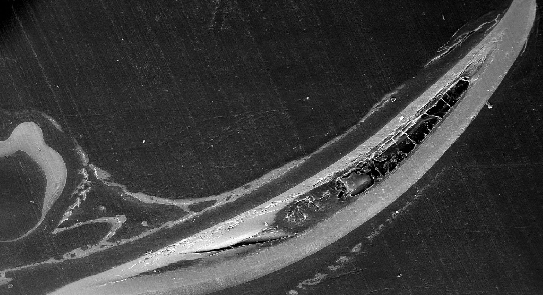
An image taken through scanning electron microscopy (SEM) shows a polished sagittal section through a mouse mandibular incisor, showing the different mineralized tissue layers.
(Image: Courtesy of Penn Engineering Today)
In a study published in the American Chemical Society’s ACS Applied Materials & Interfaces, engineers and dentists have uncovered how teeth, as biological material, hold key information for understanding rare craniofacial disorders that develop during childhood. Kyle Vining, assistant professor in materials science and engineering (MSE) at Penn’s School of Engineering and Applied Science, and preventative and restorative science at Penn’s School of Dental Medicine, leads an interdisciplinary team. The team includes Yuchen (Tracy) Jiang, a former master’s student in MSE, Kei Katsura, a pediatric dentist and postdoctoral research scholar at Children’s Hospital of Philadelphia (CHOP) and the Institute of Translational Medicine and Therapeutics at Penn, and Elizabeth Bhoj, assistant professor of pediatrics in Penn’s Perelman School of Medicine and the Division of Human Genetics at CHOP.
“People often assume that if you understand bone, you understand teeth,” says Vining. “But that’s not necessarily the case. Teeth have a different composition, require different analytical tools and behave differently during development.”
The project is centered on a deceptively simple question: “How do teeth mineralize?” To answer this question, researchers borrowed a surprising tool from geology: the nanoindenter, a device traditionally used to test the hardness of rocks. Using nanoindentation, scanning electron microscopy, energy dispersive spectroscopy (EDS), and Raman spectroscopy, the team measured everything from tooth enamel’s elasticity and stiffness to mineral contents.
While Jiang and Vining analyzed the physical properties of teeth, Katsura brought in the biological side: mouse models of Mendelian genetic disorders, many of which mimic the human versions of craniofacial syndromes. Bhoj provided further expertise on these rare diseases, helping guide the project toward real-world clinical applications.
This work is already informing the team’s future studies on genetic craniofacial diseases in mice. Long term, the researchers envision their tools used in dental clinics to screen for enamel defects, assess treatment outcomes, or even predict disease risk.
Read more at Penn Engineering Today.