We constructed epitaxial heterostructures by depositing Sr2IrO4 epitaxial thin films on ruthenates single crystals (Fig. 1) using pulsed laser deposition21,22. To minimize the potential influence of spin-spin interactions, we selected ruthenates with paramagnetic characteristics. Sr2RuO4 single crystals exhibit a tetragonal crystal structure and metallic transport behavior, while Ca3Ru1.98Ti0.02O7 single crystals are orthorhombic and undergo a metal-insulator transition at 55 K, exhibiting metallic behavior above this temperature and insulating behavior below it23,24. This transition is accompanied by minor changes in the b- and c-lattice constants (Supplementary Fig. 1). To systematically investigate the strain effects, we also studied Sr2IrO4 thin films deposited on insulating (LaAlO3)0.3(Sr2TaAlO6)0.7 (LSAT) substrates, which has a similar lattice mismatch with Sr2IrO4 as Sr2RuO4.
Fig. 1: Sr2IrO4 thin-film heterostructure systems.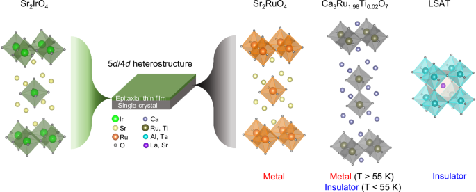
Schematic representation of Sr2IrO4 thin films interfaced with various substrates, including ruthenates single crystals and reference LSAT substrates. Sr2RuO4 and LSAT single crystals have comparable in-plane lattice constants but exhibit distinct electronic properties, with Sr2RuO4 being metallic and LSAT insulating. The Ca3Ru1.98Ti0.02O7 single crystal has a temperature-dependent metal-insulator transition, exhibiting metallic behavior above 55 K and becoming insulating below this temperature.
High-resolution Z-contrast scanning transmission electron microscopy of the Sr2IrO4/Sr2RuO4 heterostructure (Supplementary Fig. 2) reveals an atomically sharp heterointerface with minimal interfacial diffusion, similar to the sharp Sr2IrO4/Ca3Ru2O7 (ref. 25) and Sr2IrO4/Ca3Ru1.98Ti0.02O7 heterointerfaces (Supplementary Fig. 3). Distinct x-ray (0 0 l)-diffraction peaks are observed for the Sr2IrO4 thin films as well as the Sr2RuO4, Ca3Ru1.98Ti0.02O7, and LSAT substrates, accompanied by interference fringes near the (0 0 12) peak (Supplementary Fig. 4a).
X-ray reciprocal space mapping confirms that the in-plane lattice of Sr2IrO4 thin films is strained to all three substrates without relaxation (Supplementary Fig. 4b, c). Notably, the Sr2IrO4 thin films on both Sr2RuO4 and LSAT experience the same amount of compressive strain of −0.51% (Supplementary Table 1). In contrast, the Sr2IrO4 thin film on Ca3Ru1.98Ti0.02O7 undergoes a more complex strain profile: −2.22% compressive strain along the a-axis and 0.33% tensile strain along the b-axis above 55 K, which increases to 2% tensile strain along the b-axis below 55 K (Supplementary Table 2).
In recent years, RIXS has emerged as a powerful tool for collecting momentum-resolved and element-specific insights into collective magnetic excitations, such as magnons and spin-orbit excitons, in transition metal oxides26,27,28,29. Figure 2a presents representative energy loss spectra of our Sr2IrO4/Sr2RuO4 heterostructure, measured with different momentum transfers in the magnetic Brillouin zone. Figure 2b provides an intensity map derived from the energy loss spectra in Fig. 2a, highlighting the key spectral features. The low-energy range (0–0.25 eV) shows a dispersive magnetic excitation (magnon), while the higher-energy range (0.30–0.90 eV) reveals a dispersive orbital excitation (spin-orbit exciton), both of which reflect the intrinsic properties of the system18,27. The presence of well-defined dispersive magnons and spin-orbit excitons underscores the high crystallinity of the Sr2IrO4/Sr2RuO4 heterostructure, further validating the quality of the epitaxial thin films used in this study.
Fig. 2: RIXS spectra of the Sr2IrO4/Sr2RuO4 heterostructure.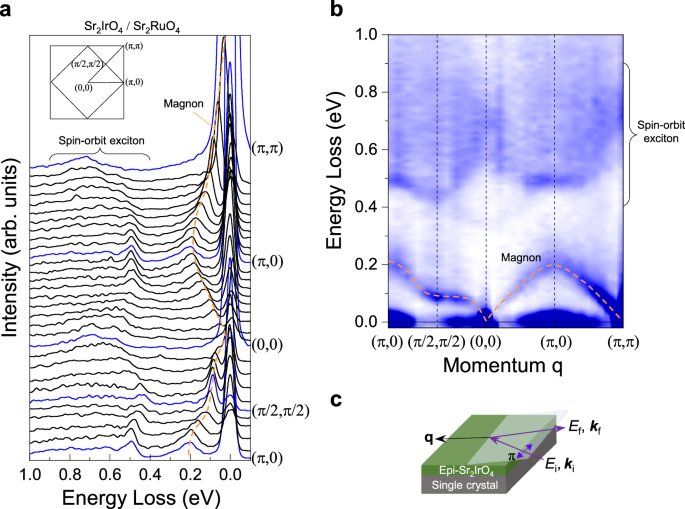
a Energy loss spectra of the Sr2IrO4/Sr2RuO4 heterostructure at 20 K with different momenta in the Brillouin zone. The inset illustrates high symmetry points of both the undistorted tetragonal unit cell and the magnetic unit cell. b Image plot of the energy loss spectra shown in (a), highlighting the dispersive magnon and spin-orbit exciton modes. The detection of well-defined dispersive features confirms the high crystalline quality of the Sr2IrO4 thin film. The orange dashed line in (a, b) serves as a guide to the magnon dispersion. The intensity scale bar is in arbitrary units. c Schematic of the horizontal scattering geometry used during the RIXS measurements.
The noteworthy feature observed in the RIXS experiments is the pronounced softening of the low-energy magnon dispersion in Sr2IrO4 films, specifically near the (π/2, π/2) zone boundary, when their interfaced single-crystal substrates transition from insulating to metallic states. This softening, by ~20 meV, is a striking contrast to the consistent magnon energies observed in other regions of the Brillouin zone. Figure 3a illustrates the low-energy magnon spectra of all Sr2IrO4 thin films along the high-symmetry directions (π, 0) and (π/2, π/2). At (π, 0), the magnon peak energy remains nearly identical, around 200 meV, across all systems studied: Sr2IrO4/Sr2RuO4, Sr2IrO4/Ca3Ru1.98Ti0.02O7 (both above and below 55 K), Sr2IrO4 /LSAT, and the Sr2IrO4 single crystal. However, at (π/2, π/2), a notable difference emerges. Thin films interfaced with metallic substrates—Sr2IrO4/Sr2RuO4 (orange) and Sr2IrO4/Ca3Ru1.98Ti0.02O7 above 55 K (red)—exhibit a significant softening of the magnon peak energy, with a reduction of approximately 20 meV. In contrast, thin films on insulating substrates, such as Sr2IrO4/Ca3Ru1.98Ti0.02O7 below 55 K (blue) and Sr2IrO4/LSAT (cyan), show magnon peak energies at (π/2, π/2) that are indistinguishable from that of the Sr2IrO4 single crystal. This shift in magnon energy exceeds the experimental error bar, underscoring the significance of this observation28.
Fig. 3: Magnon softening in Sr2IrO4 thin films interfaced with metallic substrates at the (π/2, π/2) zone boundary.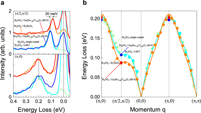
a RIXS spectra of Sr2IrO4 thin films on various substrates, compared to single-crystal Sr2IrO4 (light green) at (π, 0) and (π/2, π/2). Substrates include metallic (Sr2IrO4/Sr2RuO4 (orange) and Sr2IrO4/Ca3Ru1.98Ti0.02O7 at T > 55 K (red)) and insulating (Sr2IrO4/LSAT (cyan) and Sr2IrO4/Ca3Ru1.98Ti0.02O7 at T b Magnon dispersion extracted from the RIXS spectra and compared with single-crystal Sr2IrO4 data. The solid orange and cyan lines represent theoretical fits using the model Hamiltonian for Sr2IrO4/Sr2RuO4 and Sr2IrO4/LSAT, respectively.
Essentially, the results can be categorized into two groups (Fig. 3b): Sr2IrO4 heterostructures with insulating substrates retain higher magnon energy near (π/2, π/2), comparable to single-crystal Sr2IrO4, while those interfaced with metallic substrates exhibit a pronounced magnon energy softening, reduced by approximately 20 meV. This behavior highlights the critical role of substrate properties—particularly metallicity—in tuning the magnon dispersion of Sr2IrO4 thin films. The softened magnon energy at (π/2, π/2) is a highly unusual phenomenon, as it increases the magnon dispersion between the (π, 0) and (π/2, π/2) zone boundaries.
In typical antiferromagnetic S = 1/2 systems like La2CuO4, the magnon energy difference between these zone boundaries is relatively small, as observed in inelastic neutron scattering experiments30. However, in Sr2IrO4 single crystals (a Jeff = 1/2 pseudospin system), this energy difference is significantly larger, and it increases further in Sr2IrO4 thin films interfaced with metallic substrates. According to linear spin-wave theory18,31, the magnon energy dispersion (\({\omega }_{{{\bf{q}}}}\)) is described as: \({\omega }_{{{\bf{q}}}}=\sqrt{{A}_{{{\bf{q}}}}^{2}-{B}_{{{\bf{q}}}}^{2}}\), where: \({A}_{{{\bf{q}}}}=2({J}_{1}-{J}_{2}-{J}_{3}+{J}_{2}\cos {q}_{x}\cos {q}_{y})+ {J}_{3}(\cos {2q}_{x}+\cos 2{q}_{y})\), \({B}_{{{\bf{q}}}}={J}_{1}(\cos {q}_{x}+\cos {q}_{y})\). Here, J1, J2, and J3 represent the in-plane exchange interactions between the nearest, next-nearest, and third-nearest neighbors, respectively. The magnon energies at the (π, 0) and (π/2, π/2) zone boundaries are expressed as: \({\omega }_{\left(\pi,0\right)}\) = \(2({J}_{1}-{2J}_{2})\), \({\omega }_{\left(\frac{\pi }{2},\frac{{{\boldsymbol{\pi }}}}{{{\boldsymbol{2}}}}\right)}\) = \(2({J}_{1}-{J}_{2}-2{J}_{3})\). Thus, the experimentally observed magnon softening near the (π/2, π/2) zone boundary can be attributed to changes in J2 and J3, specifically \({2}({-}{J}_{2}+2{J}_{3})\).
By fitting the magnon dispersion data (Fig. 3b) using the Heisenberg spin model, we extracted the in-plane exchange interactions up to the fourth-nearest neighbor (J4) for the Jeff = 1/2 pseudospins in Sr2IrO4. The best-fit parameters are summarized in Supplementary Table 3: For the Sr2IrO4 thin films contacted with insulating substrates: J1 = 55 meV, J2 = −17 meV, J3 = 16 meV, and J4 = 7.3 meV. For thin films interfaced with metallic substrates: J1 = 55 meV, J2 = −21 meV, J3 = 20 meV, and J4 = 4.6 meV. Notably, while J1 remains unchanged, the magnitudes of J2 and J3 increase in Sr2IrO4 interfaced with metallic substrates, consistent with the predictions of linear spin-wave theory discussed above.
The two-magnon peak energies observed in high-resolution Raman spectra corroborate the RIXS results. Figure 4a illustrates the B2g two-magnon modes in Sr2IrO4/Sr2RuO4 and Sr2IrO4/LSAT at 10 K, while Fig. 4b presents the temperature-dependent Raman spectra of B2g two-magnon modes in Sr2IrO4/Ca3Ru1.98Ti0.02O7. The two-magnon peak energies (ω2M) were determined by fitting the data to a model function comprising two Lorentz oscillators. Notably, the two-magnon energies of thin films on metallic substrates (Sr2RuO4 and Ca3Ru1.98Ti0.02O7 above 55 K) are lower than those of thin films on insulating substrates (Ca3Ru1.98Ti0.02O7 below 55 K and LSAT). Given that the two-magnon mode primarily reflects zone boundary excitations32, this observation is consistent with the softening of the (π/2, π/2) zone boundary magnon. These results establish a strong agreement between the Raman and RIXS measurements, further confirming the substrate-dependent modification of magnon dynamics in Sr2IrO4 thin films.
Fig. 4: Softening of two-magnon modes in Sr2IrO4 thin films interfaced with metallic substrates.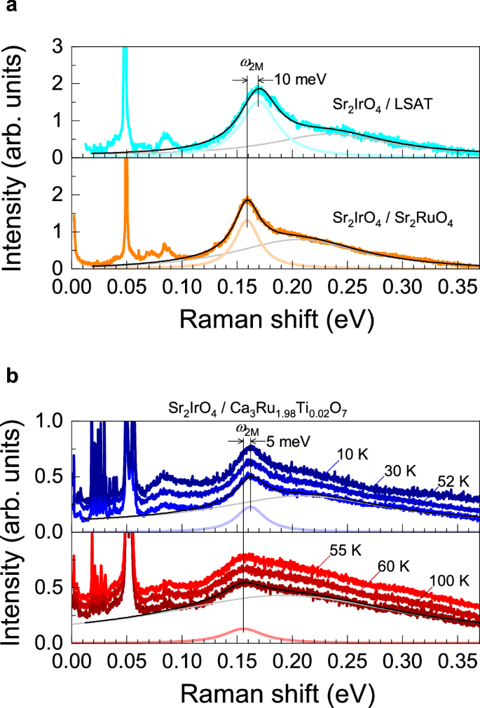
a Raman spectra of B2g two-magnon modes in Sr2IrO4/Sr2RuO4 (orange) and Sr2IrO4/LSAT (cyan) heterostructures measured at 10 K. b Temperature-dependent Raman spectra of B2g two-magnon modes in the Sr2IrO4/Ca3Ru1.98Ti0.02O7 heterostructure. The two-magnon peak energy is lower in Sr2IrO4 thin films interfaced with metallic substrates (Sr2RuO4 and Ca3Ru1.98Ti0.02O7 at T > 55 K) compared to those interfaced with insulating substrates (LSAT and Ca3Ru1.98Ti0.02O7 at T
Raman spectroscopy reveals a noticeable hardening of the phonon modes in Sr2IrO4 thin films by ~8–11 cm−1 (1–1.4 meV) when the contacted substrate transitions from insulating to metallic, likely indicating electron-phonon interactions. Figure 5a shows the A1g and B2g phonon modes of the Sr2IrO4 thin film in the Sr2IrO4/LSAT and Sr2IrO4/Sr2RuO4 heterostructures. While the phonon modes in Sr2IrO4/LSAT closely resemble those of Sr2IrO4 single crystals, the Sr2IrO4/Sr2RuO4 heterostructure exhibits a significant upward energy shift of approximately 1.4 meV for the A1g mode and 1 meV for the B2g mode. Similarly, an upward shift of up to 0.7 meV in phonon energy is observed in Sr2IrO4/Ca3Ru1.98Ti0.02O7 heterostructures when the substrate transitions from an insulating to a metallic state at 55 K (Fig. 5b).
Fig. 5: Hardening of phonons in Sr2IrO4 interfaced with metallic substrates.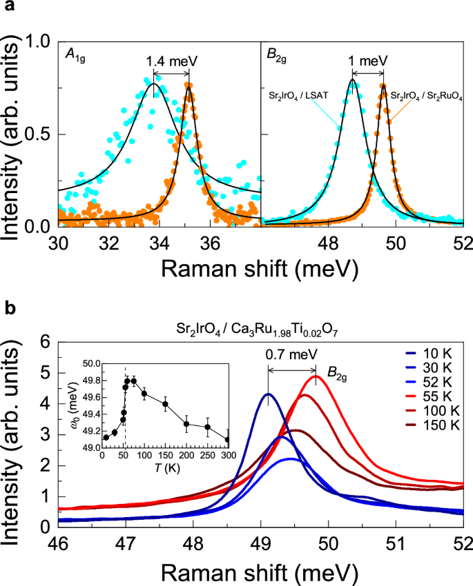
a A1g and B2g phonon modes of Sr2IrO4/Sr2RuO4 (orange) and Sr2IrO4/LSAT (cyan) heterostructures. The solid black lines represent Lorentzian fits to the data. b Temperature-dependent evolution of the B2g phonon modes in the Sr2IrO4/Ca3Ru1.98Ti0.02O7 heterostructure. Inset: Temperature-dependent peak position of the B2g phonon modes in the same heterostructure, illustrating the phonon hardening behavior as the substrate transitions from an insulating to a metallic state. Error bars represent uncertainties estimated from the peak widths and curve-fits.
It is important to note that the structural change occurring in the Ca3Ru1.98Ti0.02O7 substrate at 55 K may also contribute to the observed phonon mode shift in Sr2IrO4/Ca3Ru1.98Ti0.02O7 heterostructures. Additionally, we investigated other heterostructures, including Sr2IrO4/Ca2Ru0.91Mn0.09O4 (insulator), Sr2IrO4/Sr2RhO4 (metal), and Sr2IrO4/Ca3Ru2O7 (metal) at 10 K. These heterostructures exhibited a similar trend: the metallic substrates induced hardening of the B2g phonons in the Sr2IrO4 thin films by about 8 cm−1 (1 meV) compared to insulating substrates (Supplementary Fig. 5). Overall, a common factor across all results is the metallic state of the substrate, which strongly correlates with the observed phonon mode stiffening in Sr2IrO4 thin films.