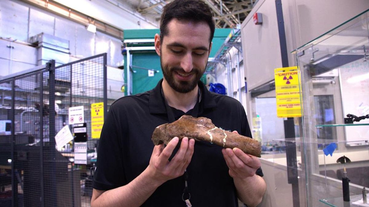
A broken rib from the largest Tyrannosaurus rex ever discovered is offering scientists a rare glimpse of a 66-million-year-old injury still in the process of healing. Researchers used neutron imaging at Oak Ridge National Laboratory to look inside the fossilized bone from a T. rex nicknamed Scotty, building detailed 3D views without damaging the preserved tissue inside.
Scotty’s rib contains a vast network of mineralized blood vessels never before observed in a fossil. Mauricio Barbi, a physics professor at the University of Regina in Saskatchewan, Canada, made that determination. Barbi called the find comparable to winning the lottery, given how rarely soft tissue like blood vessels survives the fossilization process at all.
A frozen wound
When Scotty’s rib first broke, iron-rich blood flowed into the injury site, forming new blood vessels to begin the healing process. The dinosaur died in a salty marsh before that healing finished. That marsh environment slowed decomposition enough to preserve the delicate vessel network that would otherwise have vanished entirely.

Jerit Mitchell, a University of Regina doctoral candidate in physics, leads the project under Barbi’s direction. He first discovered evidence of blood vessels in Scotty’s rib back in 2020 as an undergraduate. Working at the Canadian Light Source, Mitchell used a noninvasive X-ray technique called micro-CT scanning to confirm fossilized soft tissue survived within cut slices of the rib.
Where X-rays leave off
Neutrons and X-rays complement each other in materials science. Neutrons reveal light elements like hydrogen with unusual clarity, while X-rays excel at showing denser structures. Researchers have compared the difference to an MRI scan highlighting soft tissue, versus a standard X-ray highlighting bone.
In April 2026, the team turned to two ORNL instruments to build on their earlier X-ray findings. Those were the Multimodal Advanced Radiography Station at the High Flux Isotope Reactor and the Virtual Environment for Neutron Sciences at the Spallation Neutron Source. Marcella Berg, a University of Regina assistant professor and former ORNL postdoctoral researcher, said neutrons corroborated the team’s earlier synchrotron findings. They also added valuable new detail about soft tissue preservation, all without damaging the fossil samples themselves.
More fossils await
The two instruments serve different purposes within the same investigation. MARS produces cold neutrons well suited to revealing fine soft-tissue detail in smaller samples like bones, amber, and fossilized scales. VENUS produces higher-energy neutrons capable of generating detailed 3D images of larger specimens like Scotty’s rib.
Hassina Bilheux, lead instrument scientist for VENUS, said neutrons are often associated with battery or materials research, but prove equally valuable for investigating ancient life. Mitchell said countless fossils remain in museum collections worldwide, still holding secrets that neutron and X-ray imaging could eventually help reveal. The team plans to expand its analysis to additional fossils, comparing healing patterns and pathologies across species using the combined imaging techniques.